

Developing and testing an algorithm for automatic segmentation of the fetal face from three-dimensional ultrasound images
- PMID: 33391808
- PMCID: PMC7735327
- DOI: 10.1098/rsos.201342
Developing and testing an algorithm for automatic segmentation of the fetal face from three-dimensional ultrasound images
Abstract
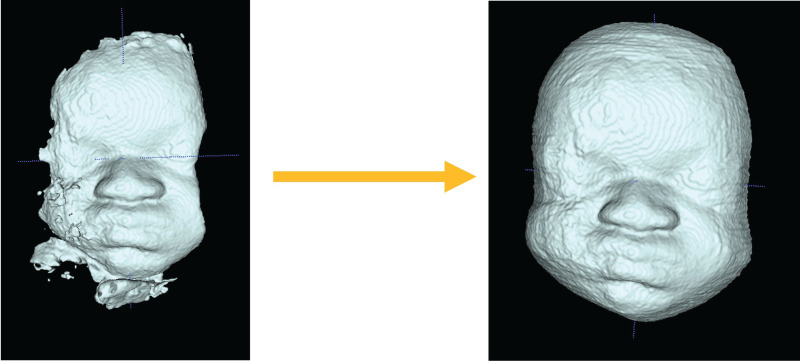
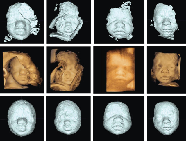
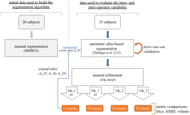
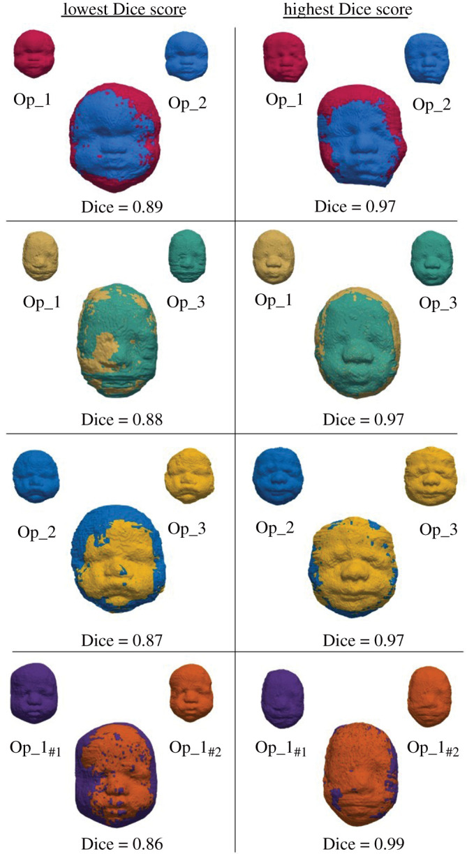
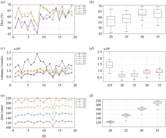
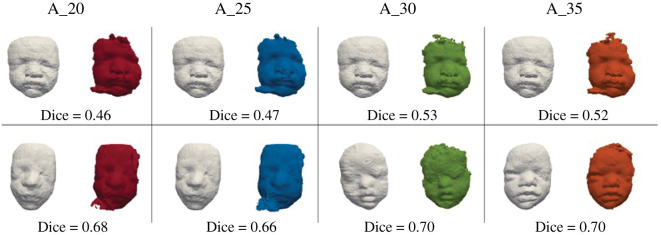
Fetal craniofacial abnormalities are challenging to detect and diagnose on prenatal ultrasound (US). Image segmentation and computer analysis of three-dimensional US volumes of the fetal face may provide an objective measure to quantify fetal facial features and identify abnormalities. We have developed and tested an atlas-based partially automated facial segmentation algorithm; however, the volumes require additional manual segmentation (MS), which is time and labour intensive and may preclude this method from clinical adoption. These manually refined segmentations can then be used as a reference (atlas) by the partially automated segmentation algorithm to improve algorithmic performance with the aim of eliminating the need for manual refinement and developing a fully automated system. This study assesses the inter- and intra-operator variability of MS and tests an optimized version of our automatic segmentation (AS) algorithm. The manual refinements of 15 fetal faces performed by three operators and repeated by one operator were assessed by Dice score, average symmetrical surface distance and volume difference. The performance of the partially automatic algorithm with difference size atlases was evaluated by Dice score and computational time. Assessment of the manual refinements showed low inter- and intra-operator variability demonstrating its suitability for optimizing the AS algorithm. The algorithm showed improved performance following an increase in the atlas size in turn reducing the need for manual refinement.
Keywords: craniofacial abnormalities; image segmentation; three-dimensional ultrasound.
© 2020 The Authors.
Conflict of interest statement
The authors declare no competing interests, financial or otherwise.
Figures
References
-
- Benoit B, Chaoui R. 2005. Three-dimensional ultrasound with maximal mode rendering: A novel technique for the diagnosis of bilateral or unilateral absence or hypoplasia of nasal bones in second-trimester screening for Down syndrome. Ultrasound Obstet. Gynecol. 25, 19–24. ( 10.1002/uog.1805) - DOI - PubMed
LinkOut - more resources
Full Text Sources
Research Materials