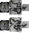
Comparison of plaque characteristics of small and large subcortical infarctions in the middle cerebral artery territory using high-resolution magnetic resonance vessel wall imaging
- PMID: 33392011
- PMCID: PMC7719918
- DOI: 10.21037/qims-20-310
Comparison of plaque characteristics of small and large subcortical infarctions in the middle cerebral artery territory using high-resolution magnetic resonance vessel wall imaging
Abstract
Background: The characteristics of plaque that ultimately lead to different subcortical infarctions remain unclear. We explored the differences in plaque characteristics between patients with small subcortical infarction (SSI) and large subcortical infarction (LSI) of the middle cerebral artery (MCA) using high-resolution magnetic resonance vessel wall imaging (HR-MRVWI).
Methods: The study group comprised 71 patients (mean age, 47.49±11.5 years; 55 male) with MCA territory ischemic stroke. Whole-brain HR-MRVWI was performed using a three-dimensional T1-weighted variable-flip-angle turbo spin echo (SPACE) sequence. Patients were divided into SSI and LSI groups based on routine MRI images. Plaque distribution was classified as the superior, inferior, ventral, or dorsal wall of the MCA. The number of quadrants with plaque formation, location of plaque, plaque burden (PB), arterial remodeling pattern (positive or negative), and degree of stenosis were analyzed and compared between groups.
Results: Of the 71 patients, 43 (60.6%) and 28 (39.4%) were identified as the SSI and LSI groups, respectively. The proportion of plaques involving only one quadrant was significantly higher in the SSI group, and these plaques were located in the superior or dorsal MCA vessel wall. There was no significant difference between groups in the proportion of plaques involving two or more quadrants, plaque distribution, or PB. Most plaques in both groups showed positive remodeling, and the percentage of remodeling pattern was similar. A significantly higher incidence of low-grade stenosis (<50%) was observed in the SSI group.
Conclusions: Both SSI and LSI may be associated with major intracranial artery atherosclerosis, but patients with SSI showed relatively fewer quadrants with plaque formation and a lesser degree of stenosis.
Keywords: Atherosclerotic plaque; ischemic stroke; magnetic resonance vessel wall imaging (MRVWI); subcortical infarction.
2021 Quantitative Imaging in Medicine and Surgery. All rights reserved.
Conflict of interest statement
Conflicts of Interest: All authors have completed the ICMJE uniform disclosure form (available at http://dx.doi.org/10.21037/qims-20-310). The authors have no conflicts of interest to declare.
Figures


References
LinkOut - more resources
Full Text Sources
Other Literature Sources
