. 2021 Jan;11(1):84-94.
doi: 10.21037/qims-20-5.

Association between anterior talofibular ligament injury and ankle tendon, ligament, and joint conditions revealed by magnetic resonance imaging

Affiliations

Association between anterior talofibular ligament injury and ankle tendon, ligament, and joint conditions revealed by magnetic resonance imaging

Israel Casado-Hernández et al. Quant Imaging Med Surg. 2021 Jan.

Abstract

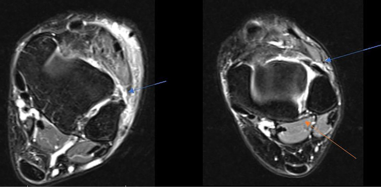
Background: The lateral ankle ligament complex is the most frequently injured ligament secondary to strong ankle inversion movement during lateral ankle sprains (LAS). Among these injuries, anterior talofibular ligament (ATFL) injury is the most frequent condition (present in 66-85% of such injuries). The purpose of this research was to use magnetic resonance imaging (MRI) to determine the association between ankle tendon, ligament, and joint conditions and ATFL injuries.

Methods: A case-control MRI study was carried out to compare the presence of ankle muscle, tendon, ligament, and joint conditions in patients with injured ATFLs (case group; n=25) and non-injured ATFLs (control group; n=25).

Results: Achilles tendinopathy was present in 1/25 (4%) patients with injured ATFLs and 7/25 (28%) non-injured ATFL subjects (P=0.048). Injured calcaneofibular ligaments (CFLs) were present in 19/25 (76%) patients with injured ATFLs and 1/25 (4%) non-injured ATFL subjects (P<0.001). Finally, injured tibiotalar joints were present in 16/25 (64%) patients with injured ATFLs and 5/25 (20%) non-injured ATFL subjects (P=0.002). Other musculoskeletal structure injuries occurred at similar rates between patients with injured ATFLs and those with non-injured ATLFs (P≥0.05).

Conclusions: Patients with ATFL injuries showed a greater presence of CFL and tibiotalar joint injuries than subjects with non-injured ATFLs.

Keywords: Ankle injuries; magnetic resonance imaging (MRI); musculoskeletal diseases; sprains and strains.


Conflict of interest statement

Conflicts of Interest: All authors have completed the ICMJE uniform disclosure form (available at http://dx.doi.org/10.21037/qims-20-5). The authors have no conflicts of interest to declare.

Figures

Figure 1
Figure 1
Axial fat-sat proton-density image. Blue star shows anterior talofibular ligament complete rupture in distal fibula portion with subcutaneous cell tissue edema.
Figure 2
Figure 2
Axial fat-sat proton-density image. Blue arrow shows peroneus brevis tendon partial rupture with a characteristic of “V” shape surrounding a thickened peroneus longus tendon. Red arrow shows fibularis head cyst lesion. Yellow arrow shows hallucis longus tendon partial injury (tenosynovitis). Orange arrow shows partial flexor digitorum tendon injury (tenosynovitis) and white arrow show partial posterior tibialis tendon injury (tenosynovitis).
Figure 3
Figure 3
Partial tibialis posterior tendon injury image at the medial part of the ankle. (A) Axial fat-sat proton-density. Blue arrow shows partial tibialis posterior tendon injury (tenosynovitis). (B) Sagittal fat-sat proton-density blue arrow shows partial tibialis posterior tendon injury (tenosynovitis).
Figure 4
Figure 4
Axial fat-sat proton-density. Orange arrows show partial tibiotalar joint injury (synovitis). White arrow shows partial calcaneofibular injury. Blue arrows show partial tibialis posterior tendon injury (tenosynovitis).
Figure 5
Figure 5
Axial fat-sat proton-density. Blue arrows show complete anterior talofibular ligament rupture. Orange arrow shows partial posterior talofibular ligament injury.
Figure 6
Figure 6
Coronal fat-sat proton-density. Yellow arrow shows partial fibulocalcaneal tendon injury.
Figure 7
Figure 7
Axial fat-sat proton-density. White arrow shows spring ligament injury (thickened). Blue arrow shows flexor hallucis longus tendon injury (tenosynovitis). Red arrow shows tibialis posterior tendon injury (tenosynovitis).
Figure 8
Figure 8
Axial fat-sat proton-density. Green arrow shows partial anterior tibiofibular ligament rupture with edema.
Figure 9
Figure 9
Sagittal fat-sat proton-density. Green arrow shows tibiotalar synovitis. White arrow shows tenosynovitis of the tibialis posterior tendon. Orange arrow shows a thickened plantar fascia.
Figure 10
Figure 10
Sagittal fast spin echo T1. Red arrow shows a thickened Achilles tendon.

References

    1. Broström L. Sprained ankles. V. Treatment and prognosis in recent ligament ruptures. Acta Chir Scand 1966;132:537-50. - PubMed
    1. Broström L. Sprained ankles. VI. Surgical treatment of "chronic" ligament ruptures. Acta Chir Scand 1966;132:551-65. - PubMed
    1. Garrick JG, Requa RK. The epidemiology of foot and ankle injuries in sports. Clin Sports Med 1988;7:29-36. 10.1016/S0278-5919(20)30956-X - DOI - PubMed
    1. Kumai T, Takakura Y, Rufai A, Milz S, Benjamin M. The functional anatomy of the human anterior talofibular ligament in relation to ankle sprains. J Anat 2002;200:457-65. 10.1046/j.1469-7580.2002.00050.x - DOI - PMC - PubMed
    1. Hertel J. Functional Anatomy, Pathomechanics, and Pathophysiology of Lateral Ankle Instability. J Athl Train 2002;37:364-75. - PMC - PubMed

LinkOut - more resources