

Titanium-Alloy Anchoring System as a Suitable Method of Extracapsular Repair
- PMID: 33392286
- PMCID: PMC7773701
- DOI: 10.3389/fvets.2020.592742
Titanium-Alloy Anchoring System as a Suitable Method of Extracapsular Repair
Abstract
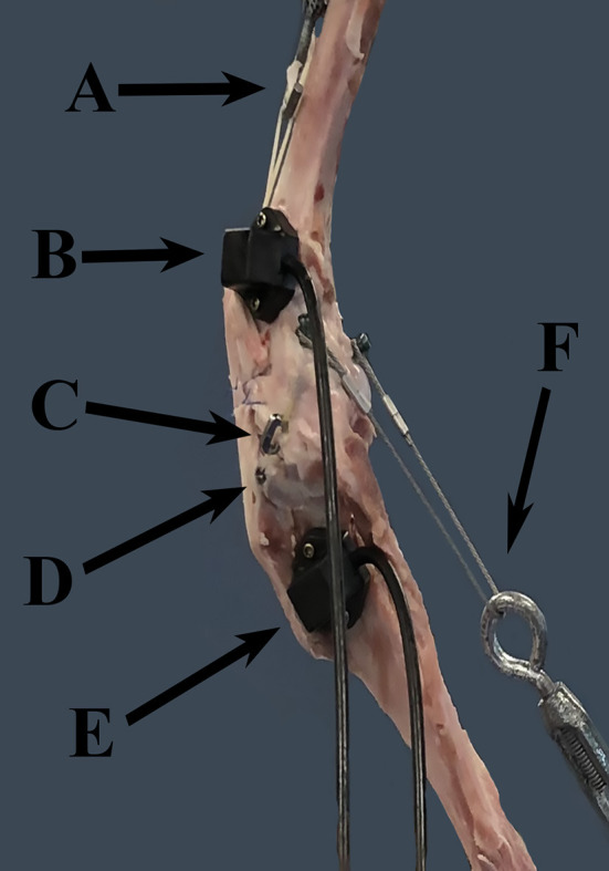
Objective: To characterize the effect of a titanium-alloy anchoring system (TAS) on the motion of the cranial cruciate ligament (CrCL) deficient stifle. To compare the motion with the TAS to that of the CrCL-intact and CrCL-deficient stifle. Study Design: Each canine pelvic limb was mounted in a loading jig under 30% body weight. Motion data was collected using an electromagnetic tracking system at stifle angles of 125°, 135°, and 145° with the CrCL-intact, CrCL-deficient and the TAS applied. Results: Total translation of the CrCL-deficient stifle following the TAS was reduced, but remained greater than the CrCL-intact stifle at angles of 125°, 135°, and 145°. Internal rotation of the TAS groups was greater than the CrCL-intact group at 145°, but not 125° and 135°. Varus motion of the TAS group was decreased compared to the CrCL-deficient group, but increased compared to the CrCL-intact group at angles of 125°, 135°, and 145°. Conclusion: Total translation and internal rotation of the CrCL-deficient stifle following the TAS differed from that of the CrCL-intact stifle. However, the TAS reduced total translation and internal rotation of the tibia relative to the femur in the CrCL-deficient stifle to levels that may yield clinically acceptable results.
Keywords: canine; cranial cruciate ligament; extracapsular; ruby; stifle.
Copyright © 2020 Dominic, Lanz, Muro, Sawyere, Aulakh, Pancotto and Seda.
Conflict of interest statement
OL is a paid consultant for KYON Veterinary Surgical Products. DS was an employee of KYON Veterinary Surgical Products. The remaining authors declare that the research was conducted in the absence of any commercial or financial relationships that could be construed as a potential conflict of interest.
Figures


References
-
- Arnoczky SP, Marshall JL. The cruciate ligaments of the canine stifle: an anatomical and functional analysis. Am J Vet Res. (1977) 38:1807–14. - PubMed
-
- Au KK, Gordon-Evans WJ, Dunning D, O'Dell-Anderson KJ, Knap KE, Griffon D, et al. Comparison of short- and long-term function and radiographic osteoarthrosis in dogs after postoperative physical rehabilitation and tibial plateau leveling osteotomy or lateral fabellar suture stabilization. Vet Surg. (2010) 39:173–80. 10.1111/j.1532-950X.2009.00628.x - DOI - PubMed
LinkOut - more resources
Full Text Sources