

Standardised computed tomographic assessment of left atrial morphology and tissue thickness in humans
- PMID: 33392384
- PMCID: PMC7772783
- DOI: 10.1016/j.ijcha.2020.100694
Standardised computed tomographic assessment of left atrial morphology and tissue thickness in humans
Abstract
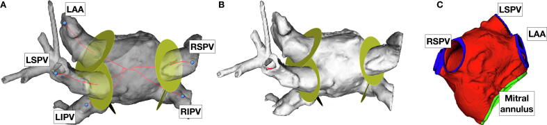
Aims: Left atrial (LA) remodelling is a common feature of many cardiovascular pathologies and is a sensitive marker of adverse cardiovascular outcomes. The aim of this study was to establish normal ranges for LA parameters derived from coronary computed tomographic angiography (CCTA) imaging using a standardised image processing pipeline to establish normal ranges in a previously described cohort.
Methods: CCTA imaging from 193 subjects recruited to the Budapest GLOBAL twin study was analysed. Indexed LA cavity volume (LACVi), LA surface area (LASAi), wall thickness and LA tissue volume (LATVi) were calculated. Wall thickness maps were combined into an atlas. Indexed LA parameters were compared with clinical variables to identify early markers of pathological remodelling.
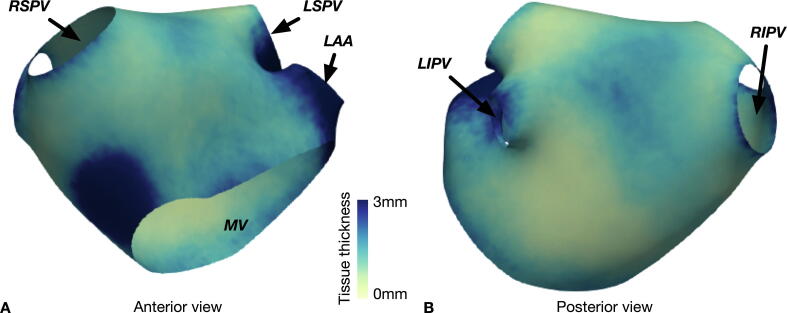
Results: LACVi is similar between sexes (31 ml/m2 v 30 ml/m2) and increased in hypertension (33 ml/m2 v 29 ml/m2, p = 0.009). LASAi is greater in females than males (47.8 ml/m2 v 45.8 ml/m2 male, p = 0.031). Median LAWT was 1.45 mm. LAWT was lowest at the inferior portion of the posterior LA wall (1.14 mm) and greatest in the septum (median = 2.0 mm) (p < 0.001). Conditions known to predispose to the development of AF were not associated with differences in tissue thickness.
Conclusions: The reported LACVi, LASAi, LATVi and tissue thickness derived from CCTA may serve as reference values for this age group and clinical characteristics for future studies. Increased LASAi in females in the absence of differences in LACVi or LATVi may indicate differential LA shape changes between the sexes. AF predisposing conditions, other than sex, were not associated with detectable changes in LAWT.Clinical trial registration:http://www.ClinicalTrials.gov/NCT01738828.
Keywords: AF, atrial fibrillation; BSA, body surface area; CCTA, cardiac computed tomography; Computed tomography (CT); DZ, dizygotic; LA, left atrium; LAA, left atrial appendage; LACV, left atrial cavity volume; LASA, left atrial surface area; LATV, left atrial tissue volume; LAWT, left atrial wall thickness; Left atrium; MZ, monozygotic; PV, pulmonary vein; Tissue thickness.
© 2020 Published by Elsevier B.V.
Conflict of interest statement
The authors report no relationships that could be construed as a conflict of interest.
Figures

Similar articles
-
Structural phenotyping in atrial fibrillation with combined cardiac CT and atrial MRI: Identifying and differentiating individual structural remodelling types in AF.J Cardiovasc Electrophysiol. 2024 Sep;35(9):1788-1796. doi: 10.1111/jce.16357. Epub 2024 Jul 4. J Cardiovasc Electrophysiol. 2024. PMID: 38965873
-
Cardiac computed tomography angiography-derived analysis of left atrial appendage morphology and left atrial dimensions for the prediction of atrial fibrillation recurrence after pulmonary vein isolation.Clin Cardiol. 2021 Nov;44(11):1636-1645. doi: 10.1002/clc.23743. Epub 2021 Oct 14. Clin Cardiol. 2021. PMID: 34651337 Free PMC article.
-
Cardiac computed tomography angiography-derived pulmonary vein volumetry as a predictor for atrial fibrillation recurrence after catheter ablation.Quant Imaging Med Surg. 2024 Mar 15;14(3):2213-2224. doi: 10.21037/qims-23-1302. Epub 2024 Mar 4. Quant Imaging Med Surg. 2024. PMID: 38545056 Free PMC article.
-
Left Atrial Wall Thickness Estimated by Cardiac CT: Implications for Catheter Ablation of Atrial Fibrillation.J Clin Med. 2024 Sep 11;13(18):5379. doi: 10.3390/jcm13185379. J Clin Med. 2024. PMID: 39336866 Free PMC article.
-
Three-dimensional atrial wall thickness maps to inform catheter ablation procedures for atrial fibrillation.Europace. 2016 Mar;18(3):376-83. doi: 10.1093/europace/euv073. Epub 2015 Apr 4. Europace. 2016. PMID: 25842272 Free PMC article.
Cited by
-
Esophageal Protection Strategies for Ablation of Atrial Fibrillation: Comparative Results of Consecutive Endoscopic Evaluation.Arq Bras Cardiol. 2025 Mar;122(3):e20230913. doi: 10.36660/abc.20230913. Arq Bras Cardiol. 2025. PMID: 40136157 Free PMC article. English, Portuguese.
-
Advancing clinical translation of cardiac biomechanics models: a comprehensive review, applications and future pathways.Front Phys. 2023 Nov 14;11:1306210. doi: 10.3389/fphy.2023.1306210. Front Phys. 2023. PMID: 38500690 Free PMC article.
-
Box Lesion Isolation of the Left Atrial Posterior Wall with Radiofrequency Ablation Restricted in Predetermined Lines for the Treatment of Persistent Atrial Fibrillation: The Prognostic Role of Acute Interventional Outcome and Trigger Identification.J Innov Card Rhythm Manag. 2023 Nov 15;14(11):5642-5653. doi: 10.19102/icrm.2023.14115. eCollection 2023 Nov. J Innov Card Rhythm Manag. 2023. PMID: 38058389 Free PMC article.
-
Left atrial-left ventricular angle, a new measure of left atrial and left ventricular remodeling.Int J Cardiovasc Imaging. 2022 Feb;38(2):435-445. doi: 10.1007/s10554-021-02411-z. Epub 2021 Sep 22. Int J Cardiovasc Imaging. 2022. PMID: 34550508 Free PMC article.
-
State of the art paper: Cardiac computed tomography of the left atrium in atrial fibrillation.J Cardiovasc Comput Tomogr. 2023 May-Jun;17(3):166-176. doi: 10.1016/j.jcct.2023.03.002. Epub 2023 Mar 23. J Cardiovasc Comput Tomogr. 2023. PMID: 36966040 Free PMC article. Review.
References
-
- Klabunde RE. CV Physiology. 2nd ed. Lippincott Williams and Wilkins; 2012. https://www.cvphysiology.com/Heart Failure/HF008. Accessed October 21, 2018.
-
- Lang R.M., Badano L.P., Mor-Avi V. Recommendations for cardiac chamber quantification by echocardiography in adults: An update from the American society of echocardiography and the European association of cardiovascular imaging. Eur Heart J Cardiovasc Imaging. 2015;16(3):233–271. doi: 10.1093/ehjci/jev014. - DOI - PubMed
-
- Nagueh S.F., Smiseth O.A., Appleton C.P. Recommendations for the Evaluation of Left Ventricular Diastolic Function by Echocardiography: An Update from the American Society of Echocardiography and the European Association of Cardiovascular Imaging. J Am Soc Echocardiogr. 2016;29(4):277–314. doi: 10.1093/ehjci/jew082. - DOI - PubMed
Associated data
Grants and funding
LinkOut - more resources
Full Text Sources
Other Literature Sources
Medical
