. 2020 Sep 30;5(1):ysaa017.
doi: 10.1093/synbio/ysaa017. eCollection 2020.

Elucidation and refinement of synthetic receptor mechanisms

Affiliations

Elucidation and refinement of synthetic receptor mechanisms

Hailey I Edelstein et al. Synth Biol (Oxf). .

Abstract

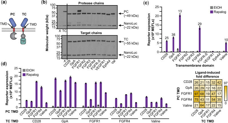
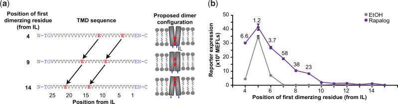
Synthetic receptors are powerful tools for engineering mammalian cell-based devices. These biosensors enable cell-based therapies to perform complex tasks such as regulating therapeutic gene expression in response to sensing physiological cues. Although multiple synthetic receptor systems now exist, many aspects of receptor performance are poorly understood. In general, it would be useful to understand how receptor design choices influence performance characteristics. In this study, we examined the modular extracellular sensor architecture (MESA) and systematically evaluated previously unexamined design choices, yielding substantially improved receptors. A key finding that might extend to other receptor systems is that the choice of transmembrane domain (TMD) is important for generating high-performing receptors. To provide mechanistic insights, we adopted and employed a Förster resonance energy transfer-based assay to elucidate how TMDs affect receptor complex formation and connected these observations to functional performance. To build further insight into these phenomena, we developed a library of new MESA receptors that sense an expanded set of ligands. Based upon these explorations, we conclude that TMDs affect signaling primarily by modulating intracellular domain geometry. Finally, to guide the design of future receptors, we propose general principles for linking design choices to biophysical mechanisms and performance characteristics.

Keywords: biosensor; cell therapy; mammalian synthetic biology; receptor engineering; transmembrane domain.


Figures

Fig. 1
Fig. 1
Protease chain tuning to improve MESA receptor performance. (a) This schematic depicts the MESA signaling mechanism. Ligand-induced receptor dimerization results in TEVp-mediated trans-cleavage to release a TF, which then enters the nucleus and induces target gene expression. Ligand-independent (background) receptor interactions can also result in TF release. (b, c) This proposed TEVp auto-inhibition strategy (b) was explored by functional evaluation (c) of MESA receptor variants in which a peptide—either a modified auto-inhibitory peptide (AIP: ELVYSQX) or protease recognition sequence (PRS: ENLYFQX), where X is a variable amino acid (e.g. AIPM: ELVYSQM)—has been appended onto the C-terminus of the protease chain (PC) TEVp. The leftmost condition is the base case (no peptide appended). Each condition uses a target chain (TC) with M at the P1’ site of the PRS. Numbers above bars indicate fold difference in reporter signal between samples treated with rapalog (dissolved in EtOH) versus EtOH (vehicle-only control). Fold difference values are shown for samples in which ligand treatment induced significant signal above background (two-way ANOVA, P < 0.05). (d, e) Juxtamembrane cleavage of the PC (d) is suggested by western blot analysis (e) of PCs tagged with 3x-FLAG on either the N-terminus or C-terminus; the PC appears to be cleaved into two fragments having sizes consistent with cleavage near the transmembrane domain (N = N-terminal product, C = C-terminal product). PCs with the TEVpD81N mutation, which renders TEVp catalytically inactive (44), were cleaved similarly to the catalytically active PCs; thus we conclude that the observed cleavage can be attributed to endogenous cellular processes. (f) PCIL substitution generally led to decreased receptor performance through increased background and/or decreased induced signal (two-way ANOVA, P < 0.05). Fold difference values are shown above bars for samples in which ligand treatment induced significant signal above background (two-way ANOVA, P < 0.05). Bars depict the mean of three biological replicates, and error bars represent the S.E.M. Outcomes from ANOVA and Tukey’s HSD tests for (c, f) are in Supplementary Notes S1 and S2. In this experiment and subsequent experiments, we employ a rapamycin analog (rapalog) as a model ligand, which induces higher ligand-induced reporter expression than does rapamycin (Supplementary Figure S4a).
Fig. 2
Fig. 2
TMD contributions to MESA receptor signaling. (a) This schematic identifies the design choice examined here—the TMD sequence. (b) Effect of TMD choice on the expression of expected bands (PC, TC, cotransfected NanoLuc loading control) versus cleavage products (CD28 and FGFR1 cases). For this experiment, chain expression levels were first normalized to that of CD28-TMD TC expression (M, upper panel) by varying transfected plasmid dose through iterative western blot analyses (Supplementary Figure S6). The X denotes a vector-only negative control (including NanoLuc); TC denotes a CD28-TMD TC. (c) Paired TMD substitution conferred varying effects on receptor performance. Labels indicate the TMD that was used on both the TC and PC. Numbers above bars indicate fold difference when the ligand induced a significant signal above background (two-way ANOVA, P < 0.05). (d) Combinatorial TMD substitution further improved receptor performance. Fold difference is reported in the heatmap at right. All combinations exhibit a significant increase in reporter expression upon ligand treatment (three-way ANOVA, P < 0.001). Bars depict the mean of three biological replicates, and error bars represent the S.E.M. Outcomes from ANOVAs and Tukey’s HSD tests for (c, d) are in Supplementary Notes S1, S2, and S3.
Fig. 3
Fig. 3
Development of a flow cytometric FRET approach to probe receptor chain association. (a) This schematic illustrates our strategy for quantifying ligand-independent (left) and ligand-mediated (right) receptor associations using Förster resonance energy transfer (FRET). Rapamycin-sensing MESA receptor ICDs were replaced with mCerulean (donor) and mVenus (acceptor) fluorophores. (b) Single-fluorophore samples were used to linearly compensate bleed-through from individual fluorophores into both the other fluorophore channel and the FRET channel. These plots also illustrate the gating used to identify cells expressing both the donor and acceptor fluorophores (mCerulean+/mVenus+). mC, mCerulean; mV, mVenus. (c) Cytosolically expressed control constructs that are expected to display low FRET (separate soluble donor and acceptor proteins) or high FRET (donor–acceptor fusion protein) differ by a vertical shift in fluorescence in the FRET channel. Fluorescence in the FRET channel is linearly correlated with donor and acceptor fluorescence, respectively. The cells shown are singlets that are transfected (miRFP670+) and that express the donor and acceptor (mCerulean+/mVenus+). (d) When FRET fluorescence is normalized to donor and acceptor fluorescence intensities by the calculated NFRET metric (equation shown), the cytosolic controls still display a vertical shift in NFRET, but NFRET only has a low correlation with donor and acceptor fluorescence, respectively; NFRET is more independent of expression differences observed across the cell population (compared to FRET fluorescence intensity in c). The NFRET metric better distinguishes low and high FRET controls than does unprocessed FRET fluorescence. The cells shown are singlets that are transfected (miRFP670+) and that express the donor and acceptor (mCerulean+/mVenus+). Experiments were conducted in biological triplicate, and individual representative samples are shown. Adjunct histograms represent probability density and are scaled to unit area. Data were analyzed as described in Section 2.
Fig. 4
Fig. 4
Effect of TMD choice on receptor chain association. (a) Receptor pairs with matched TMDs exhibit a ligand-induced increase in NFRET (27 h incubation, 100 nM rapalog) (two-way ANOVA, *** P < 0.001) (left). The CD28-TMD matched receptor pair exhibits slightly higher NFRET in the absence of ligand compared to the GpA-TMD pair and FGFR4-TMD pair (two-way ANOVA, *** P < 0.001). Fractional change in NFRET upon ligand treatment (ligand-induced NFRET fold difference) is comparable across TMDs (two-tailed Welch’s t-test, all P > 0.05) (right). In all cases, the donor fluorophore is on the FKBP chain and the acceptor fluorophore is on the FRB chain. (b) NFRET induction varies with rapalog dose (measurement at 27 h incubation). (c) Pairs of receptors with mixed and matched TMDs exhibit a significant ligand-induced increase in NFRET (27 h incubation, 100 nM rapalog) (three-way ANOVA, P < 0.001). The ligand-induced NFRET increase is comparable across mixed and matched TMD pairs. (d) Dynamics of NFRET response to ligand. By 3 h post-ligand treatment, the NFRET increase is nearly maximal (87% relative to the NFRET at 27 h). Rapa, rapalog. (e, f) In a cold (non-fluorescent) chain competition assay with matched TMD fluorescent receptors, CD28-TMD exhibited a slightly higher propensity to associate: the NFRET decrease conferred by introduction of a CD28-TMD cold chain was greater in the CD28-TMD matched case than in other cases (two-tailed Welch’s t-test, all P > 0.05 except for comparison between CD28-mediated and GpA-mediated disruption of CD28 FRET and CD28-mediated versus FGFR4-mediated disruption of CD28 FRET, * P < 0.05, ** P < 0.01). All chains contain the same ECD (FRB). Experiments were conducted in biological triplicate, and data were analyzed as described in Section 2. Error bars represent the S.E.M. Outcomes from ANOVAs and Tukey’s HSD tests for (ac) are in Supplementary Notes S2 and S3.
Fig. 5
Fig. 5
Effects of TMD dimerization geometry on receptor signaling. (a) The schematic depicts the design of synthetic TMDs used to constrain receptor dimerization geometry. Dimerization of the valine-rich alpha helices occurs at the hydrophilic glutamic acid residues. Juxtamembrane N-terminal outer linker and C-terminal inner linker (IL) residues are shown in blue. (b) Positioning the dimerizing residues at different locations in the alpha helix conferred highly varied effects on background and ligand-induced signaling. Moving the position of the first dimerizing residue from the IL from position 4 to 5, 5 to 6, 6 to 7, 7 to 8 and 9 to 10 resulted in a significant difference in ligand-induced reporter expression (two-way ANOVA, P < 0.01). Fold difference values are shown above points where ligand addition induced reporter expression that was significantly above background. Experiments were conducted in biological triplicate, and data were analyzed as described in Section 2. Error bars represent the S.E.M. Outcomes from ANOVAs and Tukey’s HSD tests for (b) are in Supplementary Note S2.
Fig. 6
Fig. 6
Tuning an expanded panel of MESA receptor systems. (a–d) Functional assays for MESA receptors for sensing rapamycin, gibberellin, abscisic acid and GFP were constructed using the full TEVp-based trans-cleavage mechanism. Axes shown on the perimeter of the heatmaps in (ad) apply to all heatmaps in (ad). (e, f) Functional assays for MESA receptors for sensing rapamycin and GFP were constructed using a revised mechanism, including previously reported H75S/L190K mutations for tuning split TEVp reconstitution propensity (26). ECDs and extracellular linker lengths are unique to each set of ligand-binding domains (Supplementary Figures S21 and S23). Axes shown on the perimeter of the heatmaps in (e) and (f) apply to all heatmaps in (e) and (f). Heatmaps display the mean from three biological replicates of reporter expression with vehicle only (gray), reporter expression with ligand (purple) and ligand-induced fold difference (gold). Within each system, a consistent plasmid dose was used across conditions. Each panel (column) is an independent experiment, and each heatmap is internally scaled by the system. Corresponding bar graphs are in Supplementary Figures S22, S24, S27–S30. Data were analyzed as described in Section 2. Outcomes from ANOVAs and Tukey’s HSD tests are in Supplementary Notes S2 and S3.
Fig. 7
Fig. 7
Generalizing principles for receptor engineering. (a) Ligand-induced inter-chain association (NFRET fold difference) varies with ECD C-terminal distance, with a negative linear relationship (y = –0.027x + 2.9, where y is NFRET fold difference and x is C-terminal distance in Å, R2 = 0.92, two-tailed Student’s t-test, P = 0.01). The ligands are rapalog, GA3-AM, ABA and secreted VEGF. The red arrow indicates the ECD C-terminal distance—the spatial displacement between C-termini of the ligand-binding domains. (b) Validation of receptor-ligand orthogonality: substantial fold differences in NFRET were observed only when each ligand was paired with its cognate ligand-binding domains. Experiments were conducted in biological triplicate, and data were analyzed as described in Section 2. Error bars represent the S.E.M. (c) This schematic synthesizes the findings of this investigation with prior MESA receptor development (26, 28) by relating receptor design choices to the proposed biophysical consequences and performance characteristics affected.

Similar articles

Cited by

References

    1. Muldoon J.J., Donahue P.S., Dolberg T.B., Leonard J.N. (2017) Building with intent: technologies and principles for engineering mammalian cell-based therapies to sense and respond. Curr. Opin. Biomed. Eng., 4, 127–133. - PMC - PubMed
    1. Guedan S., Ruella M., June C.H. (2019) Emerging cellular therapies for cancer. Annu Rev Immunol, 37, 145–171. - PMC - PubMed
    1. Roybal K.T., Lim W.A. (2017) Synthetic immunology: hacking immune cells to expand their therapeutic capabilities. Annu Rev Immunol, 35, 229–253. - PMC - PubMed
    1. Kojima R., Aubel D., Fussenegger M. (2020) Building sophisticated sensors of extracellular cues that enable mammalian cells to work as “doctors” in the body. Cell. Mol. Life Sci, 77, 3567–3581. - PMC - PubMed
    1. Schwarz K.A., Leonard J.N. (2016) Engineering cell-based therapies to interface robustly with host physiology. Adv. Drug Deliv. Rev., 105, 55–65. - PMC - PubMed

LinkOut - more resources