. 2021 Jan;7(1):e05850.
doi: 10.1016/j.heliyon.2020.e05850. Epub 2020 Dec 26.

Atlas of ACE2 gene expression reveals novel insights into transmission of SARS-CoV-2

Affiliations

Atlas of ACE2 gene expression reveals novel insights into transmission of SARS-CoV-2

Kun Sun et al. Heliyon. 2021 Jan.

Abstract

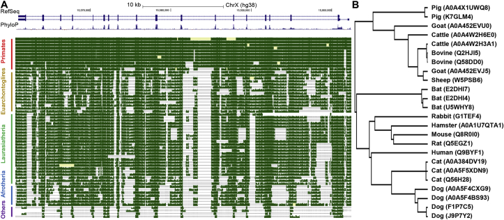
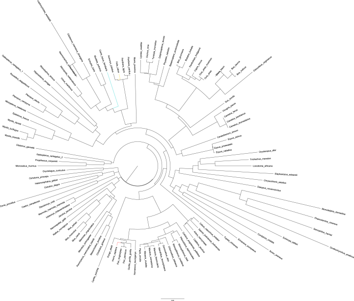
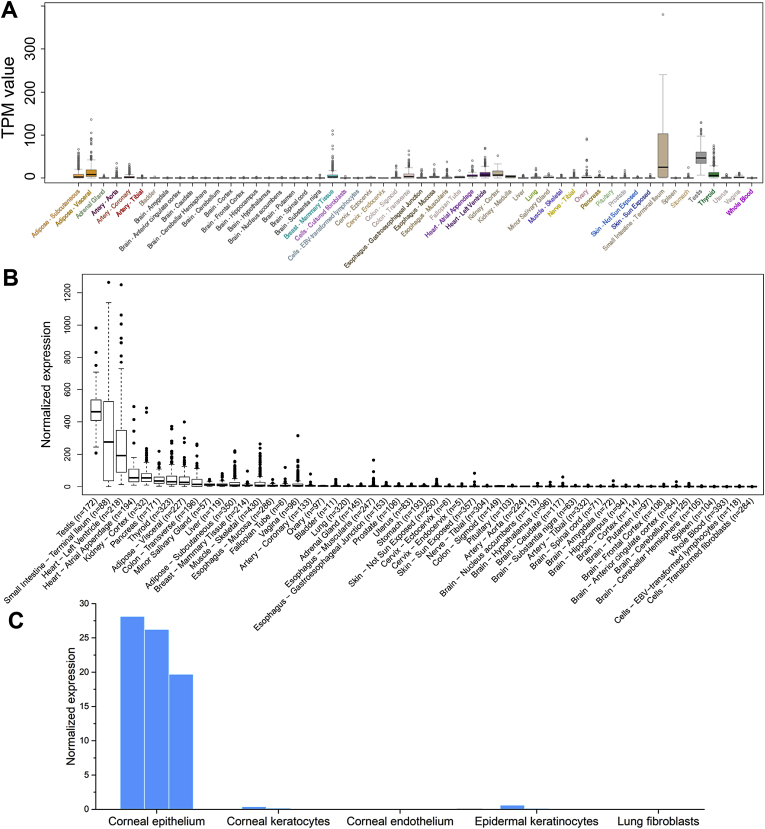
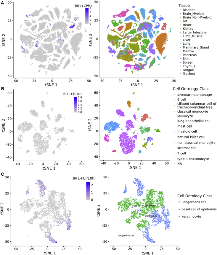
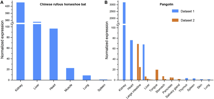
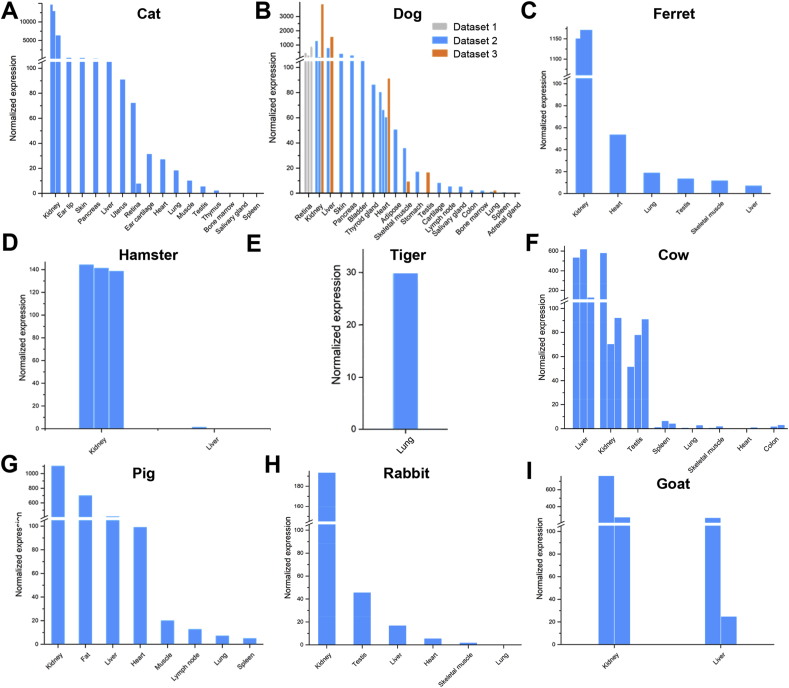
The recent pandemic, COVID-19, is caused by a novel coronavirus, SARS-CoV-2, with elusive origin. SARS-CoV-2 infects mammalian cells via ACE2, a transmembrane protein. Therefore, the conservation and expression patterns of ACE2 may provide valuable insights into tracing the carriers of SARS-CoV-2. In this work, we analyzed the conservation of ACE2 and its expression pattern among various mammalian species that are close to human beings. We show that mammalian ACE2 gene is deeply conserved at both DNA and peptide levels, suggesting that a broad range of mammals can potentially host SARS-CoV-2. We further report that ACE2 expression in certain human tissues are consistent with clinical symptoms of COVID-19 patients. Furthermore, we have built the first atlas of ACE2 expression in various common mammals, which shows that ACE2 expresses in mammalian tissues in a species-specific manner. Most notably, we observe exceptionally high expression of ACE2 in external body parts of cats and dogs, suggesting that these household pet animals could be vulnerable to viral infections and/or may serve as intermediate hosts, thus yielding novel insights into the transmission of SARS-CoV-2.

Keywords: 2019-nCov; COVID-19; Novel coronavirus; Potential host; Susceptibility.


Conflict of interest statement

The authors declare no conflict of interest.

Figures

Figure 1
Figure 1
Conservation analysis of ACE2 gene. (A) Snapshot from the UCSC genome browser showing the DNA level conservation of ACE2 gene. Horse was removed due to lack of pair-wise alignment data. (B) Phylogenetic tree based on mammal ACE2 protein sequences from UniProt database. The top panel in (A) shows RefSeq annotation of ACE2 gene in human (which contains two isoforms, and the bars stand for exons), the middle panel shows conservation scores among 100 vertebrates, higher values denote higher levels of conservation, and the best-in-genome pairwise alignments among 42 mammals are plotted in the bottom panel. For (B), the protein accession number in UniProt are shown in parentheses and some species have multiple records.
Figure 2
Figure 2
Phylogenetic tree based on mammal ACE2 protein sequences from OrthoDB database. Human, mouse, cat, and dog species are highlighted.
Figure 3
Figure 3
Expression of ACE2 gene in human tissues. (A) original TPM (Transcripts Per Kilobase of exon model per Million mapped reads) values, and (B) after normalization in GTEx project. (C) conjunctival and corneal tissues, epidermal keratinocytes, and lung fibroblasts. Available replicate experiments for tissues in panel C were shown in multiple bars.
Figure 4
Figure 4
Expression of ACE2 in murine tissues. The data was obtained from Tabula Muris project (the FACS protocol subset). (A) Expression in all cells (left) and corresponding tissue origin of these cells (right). Note that the cells were clustered using t-SNE (t-distributed Stochastic Neighbor Embedding) algorithm based on their transcriptome. (B) Expression in lungs and (C) tongue.
Figure 5
Figure 5
Expression of ACE2 gene in suspected intermediate host species. Expression levels in (A) bat, and (B) pangolin tissues. Two datasets for pangolin tissues were collected and labelled with different colors. Replicate experiments for some tissues were available and are shown as multiple bars.
Figure 6
Figure 6
Expression of ACE2 gene in common pets and livestock. Expression levels in (A) cat, (B) dog, (C) ferret, (D) pig, (E) cow, (F) rabbit, (G) hamster, (H) goat, and (I) tiger tissues. Three datasets for dog tissues were collected and labelled with different colors. Replicate experiments for some tissues were available and are shown as multiple bars.
Figure 7
Figure 7
Correlation of the two normalization methods on the Cat dataset.

References

    1. Coronaviridae Study Group of the International Committee on Taxonomy of, V The species Severe acute respiratory syndrome-related coronavirus: classifying 2019-nCoV and naming it SARS-CoV-2. Nat. Microbiol. 2020 Apr;5(4):536–544. - PMC - PubMed
    1. Zhou P. A pneumonia outbreak associated with a new coronavirus of probable bat origin. Nature. 2020 Mar;579(7798):270–273. - PMC - PubMed
    1. Letko M., Marzi A., Munster V. Functional assessment of cell entry and receptor usage for SARS-CoV-2 and other lineage B betacoronaviruses. Nat. Microbiol. 2020 Apr;5(4):562–569. - PMC - PubMed
    1. Zou X. Single-cell RNA-seq data analysis on the receptor ACE2 expression reveals the potential risk of different human organs vulnerable to 2019-nCoV infection. Front. Med. 2020 Apr;14(2):185–192. - PMC - PubMed
    1. Zhang H. Digestive system is a potential route of COVID-19: an analysis of single-cell coexpression pattern of key proteins in viral entry process. Gut. 2020;69(6):1010–1018.