

Canine models of spine disorders
- PMID: 33392448
- PMCID: PMC7770205
- DOI: 10.1002/jsp2.1109
Canine models of spine disorders
Abstract
Neck and low back pain are common among the adult human population and impose large social and economic burdens on health care and quality of life. Spine-related disorders are also significant health concerns for canine companions with etiopathogeneses, clinical presentations, and diagnostic and therapeutic options that are very similar to their human counterparts. Historically, induced and spontaneous pathology in laboratory rodents, dogs, sheep, goats, pigs, and nonhuman primates have been used for study of human spine disorders. While each of these can serve as useful preclinical models, they all have inherent limitations. Spontaneously occurring spine disorders in dogs provide highly translatable data that overcome many of the limitations of other models and have the added benefit of contributing to veterinary healthcare as well. For this scoping review, peer-reviewed manuscripts were selected from PubMed and Google Scholar searches using keywords: "intervertebral disc," "intervertebral disc degeneration," "biomarkers," "histopathology," "canine," and "mechanism." Additional keywords such as "injury," "induced model," and "nucleus degeneration" were used to further narrow inclusion. The objectives of this review were to (a) outline similarities in key features of spine disorders between dogs and humans; (b) describe relevant canine models; and (c) highlight the applicability of these models for advancing translational research and clinical application for mechanisms of disease, diagnosis, prognosis, prevention, and treatment, with a focus on intervertebral disc degeneration. Best current evidence suggests that dogs share important anatomical, physiological, histological, and molecular components of spinal disorders in humans, such that induced and spontaneous canine models can be very effective for translational research. Taken together, the peer-reviewed literature supports numerous advantages for use of canine models for study of disorders of the spine when the potential limitations and challenges are addressed.
Keywords: canine research models; intervertebral disc degeneration; spine pathology; spine‐related disorders.
© 2020 The Authors. JOR Spine published by Wiley Periodicals LLC on behalf of Orthopaedic Research Society.
Conflict of interest statement
The following authors have the following declarations: Naomi N. Lee: No conflicts to declare; Jacob S. Kramer: No conflicts to declare; Aaron M. Stoker: Arthrex, Inc: IP royalties; Other financial or material support; Musculoskeletal Transplant Foundation: IP royalties; Chantelle C. Bozynski: No conflicts to declare; Cristi R. Cook: Arthrex, Inc: IP royalties; Paid consultant; Paid presenter or speaker; Research support CONMED Linvatec: IP royalties; Paid consultant; Paid presenter or speaker Musculoskeletal Transplant Foundation: IP royalties; Paid presenter or speaker Zimmer: Research support; James T. Stannard: No conflicts to declare; Theodore J. Choma: AO Spine North America: Board or committee member Gentis, Inc: Stock or stock Options North American Spine Society: Board or committee member Scoliosis Research Society: Board or committee member; James L. Cook: Artelon: Paid consultant Arthrex, Inc: IP royalties; Paid consultant; Paid presenter or speaker; Research support AthleteIQ: IP royalties ConforMIS: Research support CONMED Linvatec: Paid consultant Coulter Foundation: Research support DePuy Synthes, A Johnson & Johnson Company: Research support Eli Lilly: Paid consultant; Research support Journal of Knee Surgery: Editorial or governing board Merial: Research support Midwest Transplant Network: Board or committee member Musculoskeletal Transplant Foundation: Board or committee member; IP royalties; Research support National Institutes of Health (NIAMS & NICHD): Research support Purina: Research support Schwartz Biomedical: Paid consultant Thieme: Publishing royalties, financial or material support Trupanion: Paid consultant U.S. Department of Defense: Research support Zimmer‐Biomet: Research support. Note: Authors James L. Cook and Cristi R. Cook are husband and wife.
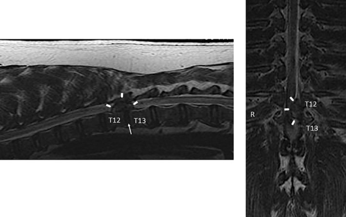
Figures


References
-
- Orakifar N, Shaterzadeh‐Yazdi MJ, Salehi R, Mehravar M, Namnik N. Muscle activity pattern dysfunction during sit to stand and stand to sit in the movement system impairment subgroups of low back pain. Arch Phys Med Rehabil. 2019;100:851‐858. - PubMed
-
- Cohen SP, Raja SN. Pathogenesis, diagnosis, and treatment of lumbar zygapophysial (facet) joint pain. Anesthesiology. 2007;106:591‐614. - PubMed
-
- Sieper J, Poddubnyy D. Axial spondyloarthritis. Lancet. 2017;390(10089):73‐84. - PubMed
-
- Schroeder GD, Kurd MF, Vaccaro AR. Lumbar spinal stenosis: how is it classified? J Am Acad Orthop Surg. 2016;24:843‐852. - PubMed
Grants and funding
LinkOut - more resources
Full Text Sources
Other Literature Sources
