

Artificial intelligence performance in detecting tumor metastasis from medical radiology imaging: A systematic review and meta-analysis
- PMID: 33392486
- PMCID: PMC7773591
- DOI: 10.1016/j.eclinm.2020.100669
Artificial intelligence performance in detecting tumor metastasis from medical radiology imaging: A systematic review and meta-analysis
Abstract
Background: Early diagnosis of tumor metastasis is crucial for clinical treatment. Artificial intelligence (AI) has shown great promise in the field of medicine. We therefore aimed to evaluate the diagnostic accuracy of AI algorithms in detecting tumor metastasis using medical radiology imaging.
Methods: We searched PubMed and Web of Science for studies published from January 1, 1997, to January 30, 2020. Studies evaluating an AI model for the diagnosis of tumor metastasis from medical images were included. We excluded studies that used histopathology images or medical wave-form data and those focused on the region segmentation of interest. Studies providing enough information to construct contingency tables were included in a meta-analysis.
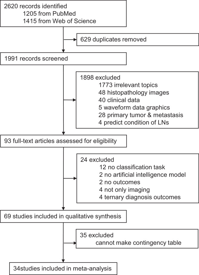
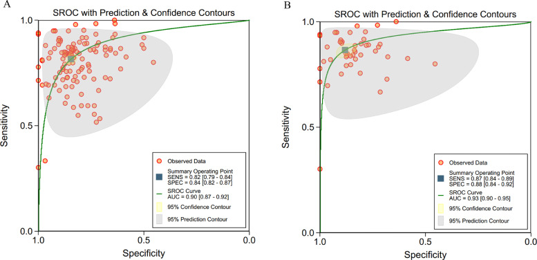
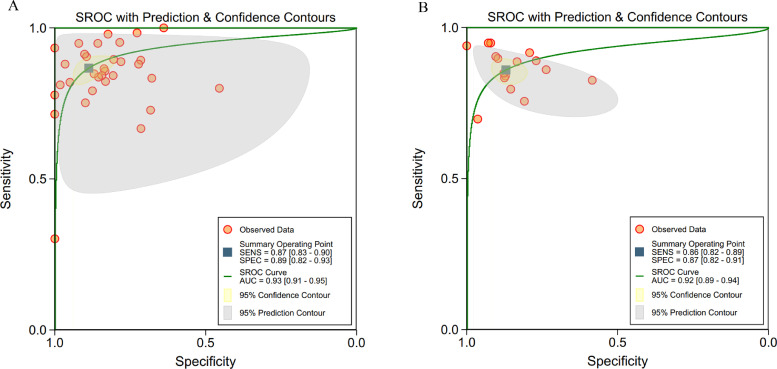
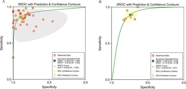
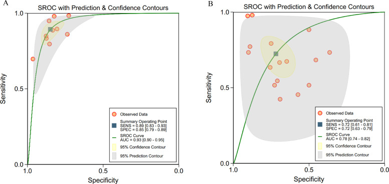
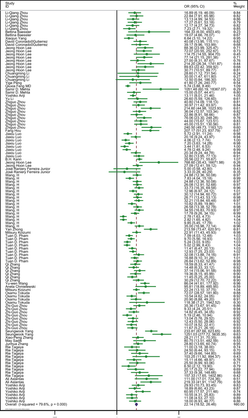
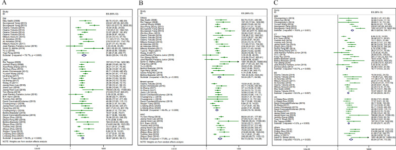
Findings: We identified 2620 studies, of which 69 were included. Among them, 34 studies were included in a meta-analysis with a pooled sensitivity of 82% (95% CI 79-84%), specificity of 84% (82-87%) and AUC of 0·90 (0·87-0·92). Analysis for different AI algorithms showed a pooled sensitivity of 87% (83-90%) for machine learning and 86% (82-89%) for deep learning, and a pooled specificity of 89% (82-93%) for machine learning, and 87% (82-91%) for deep learning.
Interpretation: AI algorithms may be used for the diagnosis of tumor metastasis using medical radiology imaging with equivalent or even better performance to health-care professionals, in terms of sensitivity and specificity. At the same time, rigorous reporting standards with external validation and comparison to health-care professionals are urgently needed for AI application in the medical field.
Funding: College students' innovative entrepreneurial training plan program .
Keywords: Artiificial intelligence; Deep learning; Diagnostic meta-analysis; Medical imaging; Tumor metastasis.
© 2020 Published by Elsevier Ltd.
Conflict of interest statement
All authors declare no competing interests.
Figures
References
-
- Yamashita K., Hosoda K., Ema A., Watanabe M. Lymph node ratio as a novel and simple prognostic factor in advanced gastric cancer. Eur J Surg Oncol. 2016;42(9):1253–1260. - PubMed
-
- Tsili A.C., Alexiou G., Naka C., Argyropoulou M.I. Imaging of colorectal cancer liver metastases using contrast-enhanced US, multidetector CT, MRI, and FDG PET/CT: a meta-analysis. Acta Radiol. 2020 284185120925481. - PubMed
LinkOut - more resources
Full Text Sources
Other Literature Sources
