

Ultrasound assessment of pulmonary fibroproliferative changes in severe COVID-19: a quantitative correlation study with histopathological findings
- PMID: 33392642
- PMCID: PMC7779089
- DOI: 10.1007/s00134-020-06328-4
Ultrasound assessment of pulmonary fibroproliferative changes in severe COVID-19: a quantitative correlation study with histopathological findings
Abstract
Purpose: This study was designed to evaluate the usefulness of lung ultrasound (LUS) imaging to characterize the progression and severity of lung damage in cases of COVID-19.
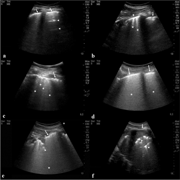
Methods: We employed a set of combined ultrasound parameters and histopathological images obtained simultaneously in 28 patients (15 women, 0.6-83 years) with fatal COVID-19 submitted to minimally invasive autopsies, with different times of disease evolution from initial symptoms to death (3-37 days, median 18 days). For each patient, we analysed eight post-mortem LUS parameters and the proportion of three histological patterns (normal lung, exudative diffuse alveolar damage [DAD] and fibroproliferative DAD) in eight different lung regions. The relationship between histopathological and post-mortem ultrasonographic findings was assessed using various statistical approaches.
Results: Statistically significant positive correlations were observed between fibroproliferative DAD and peripheral consolidation (coefficient 0.43, p = 0.02) and pulmonary consolidation (coefficient 0.51, p = 0.005). A model combining age, time of evolution, sex and ultrasound score predicted reasonably well (r = 0.66) the proportion of pulmonary parenchyma with fibroproliferative DAD.
Conclusion: The present study adds information to previous studies related to the use of LUS as a tool to assess the severity of acute pulmonary damage. We provide a histological background that supports the concept that LUS can be used to characterize the progression and severity of lung damage in severe COVID-19.
Keywords: Acute lung injury; Autopsy; COVID-19; Diffuse alveolar damage; Lung ultrasound; Minimally invasive autopsy; Pathology.
Conflict of interest statement
The authors declare that they have no conflicts of interest.
Figures


Comment in
-
Can lung ultrasound predict histologic pattern of lung injury in critically ill patients with COVID‑19?Intensive Care Med. 2021 May;47(5):629-630. doi: 10.1007/s00134-021-06372-8. Epub 2021 Feb 25. Intensive Care Med. 2021. PMID: 33630096 Free PMC article. No abstract available.
References
Publication types
MeSH terms
Grants and funding
LinkOut - more resources
Full Text Sources
Other Literature Sources
Medical
