

SARS-Cov-2 fulminant myocarditis: an autopsy and histopathological case study
- PMID: 33392658
- PMCID: PMC7779100
- DOI: 10.1007/s00414-020-02500-z
SARS-Cov-2 fulminant myocarditis: an autopsy and histopathological case study
Abstract
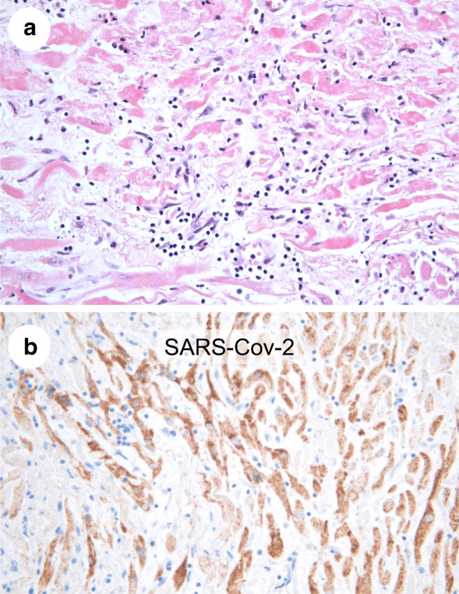
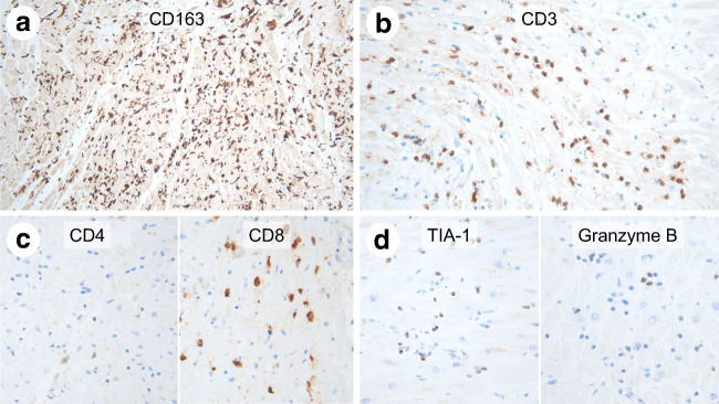
The coronavirus disease 2019 (COVID-19), due to SARS-CoV-2, is primarily a respiratory disease, causing in most severe cases life-threatening acute respiratory distress syndrome (ARDS). Cardiovascular involvement can also occur, such as thrombosis or myocarditis, generally associated with pulmonary lesions. Little is known about SARS-CoV-2-induced myocarditis. We report the case of a 69-year-old man suffering from a refractory cardiogenic shock, without significant lung involvement. Prior to death, several nasopharyngeal swabs and distal bronchoalveolar lavage were sampled in order to perform RT-PCR analyses for SARS-CoV-2-RNA, which all gave negative results. Autopsy showed coronary atherosclerosis, without acute complication. Microscopic examination of the heart revealed the existence of an intense multifocal inflammatory infiltration, in both ventricles and septum, composed in its majority of macrophages and CD8+ cytotoxic T lymphocytes (CD4/CD8 ratio: 0.11). Immunohistochemistry for anti-SARS nucleocapsid protein antibody was strongly positive in myocardial cells, but not in lung tissue. RT-PCR was realized on formalin-fixed paraffin-embedded lung and heart tissue blocks: only heart tissue was positive for SARS-CoV-2 RNA. In conclusion, this exhaustive post-mortem pathological case study of fulminant myocarditis demonstrates the presence of SARS-CoV-2 RNA in heart tissue, without significant lung involvement. Immunohistochemistry showed that the virus was specifically localized in cardiomyocytes and induced a strong cytotoxic T cells inflammatory response. This case report thus gives new insight in the pathogenesis of SARS-CoV-2-induced myocarditis and emphasizes on the importance and reliability of post-mortem analyses in order to better understand the physiopathology of this worldwide spreading new viral disease.
Keywords: Autopsy; COVID-19; Histopathology; Myocarditis; SARS-Cov-2.
Conflict of interest statement
The authors declare that they have no conflict of interest.
Figures
References
-
- Basso C, Leone O, Rizzo S, de Gaspari M, van der Wal AC, Aubry MC, Bois MC, Lin PT, Maleszewski JJ, Stone JR. Pathological features of COVID-19-associated myocardial injury: a multicentre cardiovascular pathology study. Eur Heart J. 2020;41:3827–3835. doi: 10.1093/eurheartj/ehaa664. - DOI - PMC - PubMed
-
- del Nonno F, Frustaci A, Verardo R, Chimenti C, Nicastri E, Antinori A, Petrosillo N, Lalle E, Agrati C, Ippolito G, on behalf of the INMI COVID study group (2020) Virus-negative myopericarditis in human coronavirus infection: report from an autopsy series. Circ Heart Fail 13. 10.1161/CIRCHEARTFAILURE.120.007636 - PMC - PubMed
-
- Edler C, Schröder AS, Aepfelbacher M, Fitzek A, Heinemann A, Heinrich F, Klein A, Langenwalder F, Lütgehetmann M, Meißner K, Püschel K, Schädler J, Steurer S, Mushumba H, Sperhake JP. Dying with SARS-CoV-2 infection-an autopsy study of the first consecutive 80 cases in Hamburg, Germany. Int J Legal Med. 2020;134:1275–1284. doi: 10.1007/s00414-020-02317-w. - DOI - PMC - PubMed
Publication types
MeSH terms
LinkOut - more resources
Full Text Sources
Other Literature Sources
Medical
Research Materials
Miscellaneous
