

doi: 10.1007/s11748-020-01565-2.
Epub 2021 Jan 3.
Initial experience of virtual-assisted lung mapping utilizing both indocyanine green and indigo carmine
Affiliations
- PMID: 33392864
- PMCID: PMC8131280
- DOI: 10.1007/s11748-020-01565-2
Item in Clipboard
Initial experience of virtual-assisted lung mapping utilizing both indocyanine green and indigo carmine
Gen Thorac Cardiovasc Surg.
2021 Jun.
Abstract
Virtual-assisted lung mapping is a bronchoscopic multiple dye marking technique that facilitates sublobar lung resections for unidentifiable pulmonary tumors. Marking failure reportedly occurs in 10% of cases. To overcome this limitation, we developed indocyanine green virtual-assisted lung mapping that uses indocyanine green in addition to indigo carmine. Here, we report our initial experience of indocyanine green virtual-assisted lung mapping.
Keywords: Indocyanine green; Thoracic surgery; Virtual-assisted lung mapping.
Conflict of interest statement
The authors disclose no conflict of interest for this work.
Figures
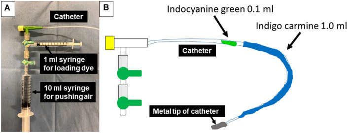
Equipment and preloading of ICG and IC. a A metal-tipped catheter was connected by syringes for loading dye and pushing air. b Then, 0.1 ml of ICG and 1.0 ml of IC were preloaded into the catheter
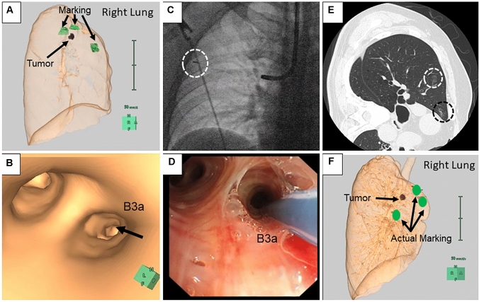
Steps involved in the ICG VAL-MAP procedure. a, b Before the bronchoscopic procedure, lung mapping was planned and a virtual bronchoscopic navigation image was created using Synapse Vincent. c X-ray fluoroscopy confirmed the tip of the catheter (surrounded by a white-dotted circle) reached the pleura. d ICG and IC dyes were injected into the targeted bronchus. e The post-VAL-MAP chest CT image shows the location of the lesion (surrounded by a white-dotted circle) and the actual marking (surrounded by a black-dotted circle). f Three-dimensional image of post-VAL-MAP chest CT was reconstructed for surgery
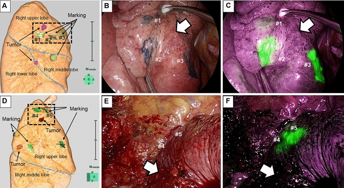
The post-VAL-MAP CT image and actual image of markings. a, b, c The post-VAL-MAP chest CT image and actual intraoperative images of markings in patient #1. IC dye markings are visible (b); however, only one ICG marking is faintly visible in marking #1 (c). The white arrows indicate the tumor location. d, e, f A post-VAL-MAP chest CT image and actual intraoperative images of markings in patient #2. IC dye markings are invisible due to tight pleural adhesion (e). However, the ICG dye markings are visible (f). The white arrows indicate the tumor location
References
MeSH terms
Substances
LinkOut - more resources
Full Text Sources
Other Literature Sources
Medical
