. 2021 Mar;26(2):417-431.
doi: 10.1007/s12192-020-01188-9. Epub 2021 Jan 4.

HSF-1 displays nuclear stress body formation in multiple tissues in Caenorhabditis elegans upon stress and following the transition to adulthood

Affiliations

HSF-1 displays nuclear stress body formation in multiple tissues in Caenorhabditis elegans upon stress and following the transition to adulthood

Andrew Deonarine et al. Cell Stress Chaperones. 2021 Mar.

Abstract

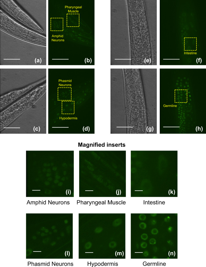
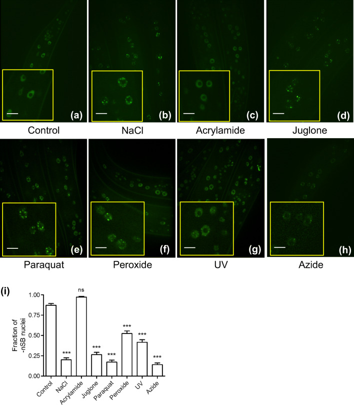
The transcription factor heat shock factor-1 (HSF-1) regulates the heat shock response (HSR), a cytoprotective response induced by proteotoxic stresses. Data from model organisms has shown that HSF-1 also has non-stress biological roles, including roles in the regulation of development and longevity. To better study HSF-1 function, we created a C. elegans strain containing HSF-1 tagged with GFP at its endogenous locus utilizing CRISPR/Cas9-guided transgenesis. We show that the HSF-1::GFP CRISPR worm strain behaves similarly to wildtype worms in response to heat and other stresses, and in other physiological processes. HSF-1 was expressed in all tissues assayed. Immediately following the initiation of reproduction, HSF-1 formed nuclear stress bodies, a hallmark of activation, throughout the germline. Upon the transition to adulthood, of HSF-1 nuclear stress bodies appeared in most somatic cells. Genetic loss of the germline suppressed nuclear stress body formation with age, suggesting that the germline influences HSF-1 activity. Interestingly, we found that various neurons did not form nuclear stress bodies after transitioning to adulthood. Therefore, the formation of HSF-1 nuclear stress bodies upon the transition to adulthood does not occur in a synchronous manner in all cell types. In sum, these studies enhance our knowledge of the expression and activity of the aging and proteostasis factor HSF-1 in a tissue-specific manner with age.

Keywords: Aging; C. elegans; Cell stress; HSF-1; Heat shock response; Nuclear stress bodies.


Conflict of interest statement

The authors declare that they have no conflict of interest.

Figures

Fig. 1
Fig. 1
The C. elegans HSF-1::GFP CRISPR model shows that HSF-1::GFP forms nuclear stress bodies (nSBs) in hypodermal cells in response to heat shock. (a, b) Expression of HSF-1::GFP from the HSF-1::GFP CRISPR strain (SDW015) grown to the L4/YA stage was analyzed by fluorescence microscopy with and without heat shock (HS) for 5 min at 33 °C. Without heat shock, HSF-1::GFP exhibits a diffuse nuclear pattern in the hypodermal cells examined. Upon heat shock, there is a redistribution of signal into nuclear stress bodies (nSBs). (c, d) Expression of HSF-1::GFP from an HSF-1::GFP MosSCI strain (OG497), with and without HS for 5 min at 33 °C. This strain is shown as a comparison to the HSF-1::GFP CRISPR strain and displays similar results. The insets represent zoomed-in images. (e) Hypodermal nuclei from SDW015 and OG497 were scored for the appearance of nSBs in A–D and the fraction of those containing no nSBs was calculated and plotted for n ≥ 8 replicate worms. Scale bar within zoomed image insert represents 5 microns. (f) CRISPR/Cas9-mediated transgenesis of the endogenous hsf-1 locus in C. elegans to include a C-terminal GFP tag. Cas9 was programmed with a guiding RNA (gRNA) to create a double-strand break upstream of the translational stop site in exon eight of HSF-1. This break was then repaired via homologous recombination utilizing unc-119 (+) as a non-fluorescent marker of repair. Green fluorescent protein (GFP) was cloned in a direct fusion to exon eight and 286 bp of the hsf-1 3’ UTR was retained following the translational stop site of GFP. Upstream and downstream homology arms (U.S. and D.S. homology) of 2.0 kb flank the targeted double-strand break location
Fig. 2
Fig. 2
The C. elegans HSF-1::GFP CRISPR model shows that HSF-1::GFP provides thermotolerance, allows the induction of hsp mRNA upon heat shock, and does not alter thermotolerance, brood size, or lifespan. (a, b) N2 (wildtype) and SDW015 (HSF-1::GFP CRISPR) animals at the L4/YA stage were subjected to a 1-h HS at 33 °C followed by immediate RNA extraction for qRT-PCR analysis of hsp-16.2 and hsp-70 (C12C8.1) expression. Three independent biological replicates were assessed in technical triplicate using the ΔΔCt analysis method. Each sample was compared to N2 (−HS) for statistical measurements. ***p value < 0.0001; ns = p value not significant. (c) Survival of N2 (wildtype) and SDW015 (HSF-1::GFP CRISPR) worm strains after being subjected to 37 °C for 2 h at the L4/YA stage. Survival was assessed 24 h post heat shock via response to gentle touch with a platinum wire. 150 worms were used for each experiment in three independent biological replicates. Results were analyzed using a two-tailed t test. The difference between the two results was not statistically significant, with a p value of 0.39. (d) RNAi targeting HSF-1::GFP alters brood size. N2 (wildtype) and SDW015 (HSF-1::GFP CRISPR) animals were grown on empty L4440 RNAi feeding vector (Control), HSF-1 RNAi, or GFP RNAi starting from L1. Total brood size of N2 (wildtype) and SDW015 (HSF-1::GFP CRISPR) animals was determined by assessing live hatched offspring measured during the first 5 days of adulthood. Eighteen worms were used for each experiment in three independent biological replicates. For significance, all samples are compared to N2 (Ctrl RNAi). ***p value < 0.0001; ns = p value not significant. (e) RNAi targeting HSF-1::GFP inhibits thermotolerance. N2 (wildtype) and SDW015 (HSF-1::GFP CRISPR) animals were grown on empty L4440 RNAi feeding vector (EV), HSF-1, or GFP RNAi starting from L1. Thermotolerance was conducted by exposure to 37 °C for 2 h at the L4/YA stage and assessing for survival 24 h later. Animals were considered alive if they responded to a gentle touch via a platinum wire. 150 worms were used for each experiment in three independent biological replicates. For significance, all samples are compared to N2 (Ctrl RNAi). **p value < 0.001; ns = p value not significant. (f) hsf-1 RNAi inhibits lifespan in N2 and in HSF-1::GFP CRISPR animals. N2 (wildtype) and SDW015 (HSF-1::GFP CRISPR) animals were grown on empty L4440 RNAi feeding vector (EV) or hsf-1 RNAi starting from L1 and measured for live/dead every other day starting at day 5 of adulthood. Worms were considered alive if they responded to a gentle touch via worm pick. Greater that 120 worms were used for each experiment, in three biological replicates. For significance, all samples are compared to N2 (Ctrl RNAi). (g) GFP RNAi inhibits lifespan in HSF-1::GFP CRISPR animals. N2 (wildtype- the same data as plotted in (F)) and SDW015 (HSF-1::GFP CRISPR) animals were grown on empty L4440 RNAi feeding vector (EV) or GFP RNAi starting from L1 and measured for live/dead every other day starting at day 5 of adulthood. Worms were considered alive if they responded to a gentle touch via worm pick. Greater that 120 worms were used for each experiment, in three biological replicates. For significance, all samples are compared to N2 (Ctrl RNAi). (h) Median survival for figures F/G plotted as bar graph. For significance, all conditions were compared to N2 (Ctrl RNAi). Significance was determined by conducting a One-Way ANOVA followed by a Tukey post hoc test comparison of all columns (D–E, H) or survival curve analysis (F–G). Significance is indicated by *p value < 0.05. “ns”: not significant
Fig. 3
Fig. 3
HSF-1::GFP CRISPR is expressed in multiple cell types and forms nuclear stress bodies in some germline cells. HSF-1::GFP from strain SDW015 grown to the L4/YA stage shows nuclear expression in the amphid neurons (A/I), pharyngeal muscle (A/J), intestine (F/K), phasmid neurons (D/L), hypodermis (D/M) and germline (H/N), as indicated. Scale bar in (ah) represents 45 microns. Scale bar in (in) represents 5 microns
Fig. 4
Fig. 4
Confocal fluorescence images show the formation of HSF-1:GFP nSBs in response to multiple cytotoxic stressors. (ah) SDW015 animals at the L4/YA stage were exposed to control conditions (a), sodium chloride at 600 mM for 30 min (b), acrylamide at 7 mM (c), juglone at 38 μM for 30 min (d), paraquat at 5 mM for 2 h (e), peroxide at 7.5 mM for 2 h (f), UV radiation at 300 J/cm2 (g), or azide at 5 mM for 5 min (h), and then assessed for the presence of nSBs in hypodermal nuclei as previously described. (I) Hypodermal nuclei were scored for the appearance of HSF-1::GFP nSBs and the fraction of cells displaying no nSBs (−nSBs) in conditions A–H was calculated and plotted for n ≥ 8 replicate worms. For significance, a One-Way ANOVA was performed with a Tukey post hoc test of all comparisons. For indicated significance, all conditions were compared to control. ***p value < 0.0001. Scale bar within zoomed image represents 5 microns
Fig. 5
Fig. 5
HSF-1::GFP forms nSBs upon transition to adulthood in hypodermal cells and germline cells, an effect that is suppressed by genetic loss of the germline. (ac) Confocal fluorescence images of HSF-1::GFP CRISPR worms (SDW015) show expression of HSF-1::GFP in hypodermal cell nuclei during the transition to adulthood in C. elegans. The formation of nSBs occurs in hypodermal cell nuclei within a 24-h window between the L4 stage (0 h) to young adulthood (YA) (~ 4–6 h post L4), and gravid adulthood (GA) (24 h post L4). (df) Confocal fluorescence images show that the formation of nSBs upon transition to adulthood is suppressed in HSF-1::GFP CRISPR animals carrying the glp-1 (e2144) mutation (strain name SDW050). (gi) Confocal fluorescence images show the robust formation of nSBs upon the transition to adulthood throughout the germline. (j) Hypodermal nuclei from HSF-1::GFP (SDW015) or HSF-1::GFP; glp-1(e2144) (SDW050) animals at the last larval stage (L4), young adult (YA), or gravid adult (GA) were scored for the appearance of HSF-1::GFP nSBs and the fraction of cells displaying no nSBs (−nSBs) was calculated and plotted for n ≥ 8 replicate worms from images (A–F). Significance indicated compares the gravid adult condition of HSF-1::GFP (SDW015) to the gravid adult condition of HSF-1::GFP; glp-1(e2144) (SDW050), *** indicates p < 0.0001. (k) HSF-1::GFP; glp-1 (e2144) (SDW050) animals were grown to the 9th day of adulthood on OP50-1 plates and hypodermal nuclei were scored for the appearance of HSF-1::GFP nSBs and the fraction of cells displaying no nSBs (−nSBs) was calculated and plotted for n ≥ 8 replicate worms per timepoint. Scale bar in zoomed-in images represents 5 microns
Fig. 6
Fig. 6
The transition to adulthood does not induce the localization of HSF-1::GFP into nuclear stress bodies in Touch Receptor Neurons (TRNs) PLM and ALM neurons, an effect that is independent of neuronal ensheathment. The Touch Receptor Neuron (TRN) co-marker pmec-17::RFP was genetically crossed into HSF-1::GFP CRISPR (SDW015) to generate HSF-1::GFP CRISPR; pmec-17::RFP (SDW077). (a) For each age, n ≥ 28 replicate animals of HSF-1::GFP CRISPR; pmec-17::RFP (SDW077) were examined and scored for the appearance of HSF-1::GFP nSBs within the PLM neuron at the L4 stage, young adult (YA), and gravid adult (GA) and the fraction of cells displaying no nSBs (−nSBs) was calculated. (b) SDW077 animals carrying the wildtype sequence variant of mec-1 or genetically crossed to the ensheathment defective mec-1 (e1292) were grown to gravid adulthood (24 hours post L4) and scored for the appearance of HSF-1::GFP nSBs within the PLM and ALM neurons and the fraction of cells displaying no nSBs (−nSBs) was calculated for n = 20 replicate individuals. (cj) Confocal images of PLM and ALM neurons as graphed in (B) scale bar represents 5 microns

Similar articles

Cited by

References

    1. Ahn SG, Thiele DJ. Redox regulation of mammalian heat shock factor 1 is essential for Hsp gene activation and protection from stress. Genes Dev. 2003;17(4):516–528. doi: 10.1101/gad.1044503. - DOI - PMC - PubMed
    1. Arantes-Oliveira N, Apfeld J, Dillin A, Kenyon C. Regulation of life-span by germ-line stem cells in Caenorhabditis elegans. Science. 2002;295(5554):502–505. doi: 10.1126/science.1065768. - DOI - PubMed
    1. Ben-Zvi A, Miller EA, Morimoto RI. Collapse of proteostasis represents an early molecular event in Caenorhabditis elegans aging. Proc Natl Acad Sci. 2009;106(35):14914–14919. doi: 10.1073/pnas.0902882106. - DOI - PMC - PubMed
    1. Berman JR, Kenyon C. Germ-cell loss extends C. elegans life span through regulation of DAF-16 by kri-1 and lipophilic-hormone signaling. Cell. 2006;124(5):1055–1068. doi: 10.1016/j.cell.2006.01.039. - DOI - PubMed
    1. Biamonti G. Nuclear stress bodies: a heterochromatin affair? Nat Rev Mol Cell Biol. 2004;5(6):493–498. doi: 10.1038/nrm1405. - DOI - PubMed

Publication types

MeSH terms

Substances