

Multiple potential roles of thymosin β4 in the growth and development of hair follicles
- PMID: 33393222
- PMCID: PMC7875905
- DOI: 10.1111/jcmm.16241
Multiple potential roles of thymosin β4 in the growth and development of hair follicles
Abstract
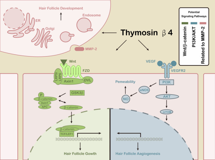
The hair follicle (HF) is an important mini-organ of the skin, composed of many types of cells. Dermal papilla cells are important signalling components that guide the proliferation, upward migration and differentiation of HF stem cell progenitor cells to form other types of HF cells. Thymosin β4 (Tβ4), a major actin-sequestering protein, is involved in various cellular responses and has recently been shown to play key roles in HF growth and development. Endogenous Tβ4 can activate the mouse HF cycle transition and affect HF growth and development by promoting the migration and differentiation of HF stem cells and their progeny. In addition, exogenous Tβ4 increases the rate of hair growth in mice and promotes cashmere production by increasing the number of secondary HFs (hair follicles) in cashmere goats. However, the molecular mechanisms through which Tβ4 promotes HF growth and development have rarely been reported. Herein, we review the functions and mechanisms of Tβ4 in HF growth and development and describe the endogenous and exogenous actions of Tβ4 in HFs to provide insights into the roles of Tβ4 in HF growth and development.
Keywords: development; growth; hair follicle; thymosin β4.
© 2021 The Authors. Journal of Cellular and Molecular Medicine published by Foundation for Cellular and Molecular Medicine and John Wiley & Sons Ltd.
Conflict of interest statement
The authors declare no conflicts of interest.
Figures
Similar articles
-
Thymosin β4 Identified by Transcriptomic Analysis from HF Anagen to Telogen Promotes Proliferation of SHF-DPCs in Albas Cashmere Goat.Int J Mol Sci. 2020 Mar 25;21(7):2268. doi: 10.3390/ijms21072268. Int J Mol Sci. 2020. PMID: 32218218 Free PMC article.
-
Role of thymosin beta 4 in hair growth.Mol Genet Genomics. 2016 Aug;291(4):1639-46. doi: 10.1007/s00438-016-1207-y. Epub 2016 Apr 29. Mol Genet Genomics. 2016. PMID: 27130465
-
Thymosin Beta-4 Induces Mouse Hair Growth.PLoS One. 2015 Jun 17;10(6):e0130040. doi: 10.1371/journal.pone.0130040. eCollection 2015. PLoS One. 2015. PMID: 26083021 Free PMC article.
-
Thymosin β4 and Actin: Binding Modes, Biological Functions and Clinical Applications.Curr Protein Pept Sci. 2023;24(1):78-88. doi: 10.2174/1389203724666221201093500. Curr Protein Pept Sci. 2023. PMID: 36464872 Review.
-
Platelet function and thymosin β4.Biol Chem. 2012 Jul;393(7):595-8. doi: 10.1515/hsz-2012-0131. Biol Chem. 2012. PMID: 22944663 Review.
Cited by
-
Screening and expression validation of key proteins for secondary hair follicle growth in cashmere goats based on iTRAQ quantitative proteomics technology.Front Vet Sci. 2024 Oct 15;11:1441074. doi: 10.3389/fvets.2024.1441074. eCollection 2024. Front Vet Sci. 2024. PMID: 39474271 Free PMC article.
-
Th22 is the effector cell of thymosin β15-induced hair regeneration in mice.Inflamm Regen. 2024 Jan 8;44(1):3. doi: 10.1186/s41232-023-00316-z. Inflamm Regen. 2024. PMID: 38191481 Free PMC article.
-
Recombinant human thymosin beta-4 (rhTβ4) improved scalp condition and microbiome homeostasis in seborrheic dermatitis.Microb Biotechnol. 2021 Sep;14(5):2152-2163. doi: 10.1111/1751-7915.13897. Epub 2021 Jul 28. Microb Biotechnol. 2021. PMID: 34318587 Free PMC article.
-
Single-Cell Sequencing Reveals Differential Cell Types in Skin Tissues of Liaoning Cashmere Goats and Key Genes Related Potentially to the Fineness of Cashmere Fiber.Front Genet. 2021 Nov 10;12:726670. doi: 10.3389/fgene.2021.726670. eCollection 2021. Front Genet. 2021. PMID: 34858469 Free PMC article.
-
SOX18 Promotes the Proliferation of Dermal Papilla Cells via the Wnt/β-Catenin Signaling Pathway.Int J Mol Sci. 2023 Nov 23;24(23):16672. doi: 10.3390/ijms242316672. Int J Mol Sci. 2023. PMID: 38068994 Free PMC article.
References
-
- Morioka K. Hair follicle: Differentiation under the electron microscope. An atlas: Springer Ebooks; 2005.
-
- Cotsarelis G, Sun TT, Lavker RM. Label‐retaining cells reside in the bulge area of pilosebaceous unit: implications for follicular stem cells, hair cycle, and skin carcinogenesis. Cell. 1990;61:1329‐1337. - PubMed
-
- Oshima H, Rochat A, Kedzia C, Kobayashi K, Barrandon Y. Morphogenesis and renewal of hair follicles from adult multipotent stem cells. Cell. 2001;104:233‐245. - PubMed
Publication types
MeSH terms
Substances
Grants and funding
LinkOut - more resources
Full Text Sources
Other Literature Sources
Research Materials
Miscellaneous
