Review
. 2021 Jan 4;131(1):e142241.
doi: 10.1172/JCI142241.

Insulin signaling in health and disease

Review

Insulin signaling in health and disease

Alan R Saltiel. J Clin Invest. .

Abstract

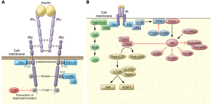
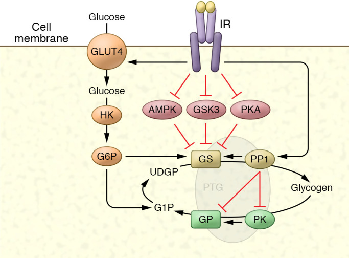
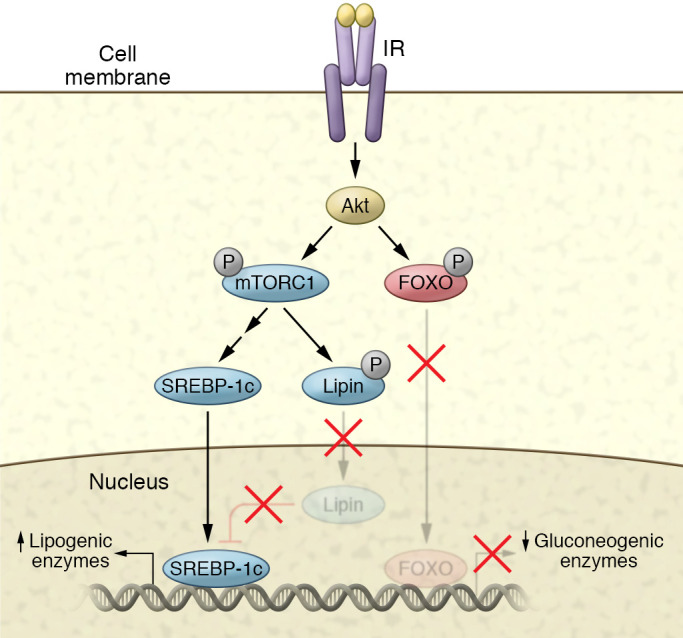
The molecular mechanisms of cellular insulin action have been the focus of much investigation since the discovery of the hormone 100 years ago. Insulin action is impaired in metabolic syndrome, a condition known as insulin resistance. The actions of the hormone are initiated by binding to its receptor on the surface of target cells. The receptor is an α2β2 heterodimer that binds to insulin with high affinity, resulting in the activation of its tyrosine kinase activity. Once activated, the receptor can phosphorylate a number of intracellular substrates that initiate discrete signaling pathways. The tyrosine phosphorylation of some substrates activates phosphatidylinositol-3-kinase (PI3K), which produces polyphosphoinositides that interact with protein kinases, leading to activation of the kinase Akt. Phosphorylation of Shc leads to activation of the Ras/MAP kinase pathway. Phosphorylation of SH2B2 and of Cbl initiates activation of G proteins such as TC10. Activation of Akt and other protein kinases produces phosphorylation of a variety of substrates, including transcription factors, GTPase-activating proteins, and other kinases that control key metabolic events. Among the cellular processes controlled by insulin are vesicle trafficking, activities of metabolic enzymes, transcriptional factors, and degradation of insulin itself. Together these complex processes are coordinated to ensure glucose homeostasis.


Conflict of interest statement

Conflict of interest: ARS is a cofounder of Elgia Therapeutics, and receives funding for his laboratory from Dong-A ST Co.

Figures

Figure 1
Figure 1. The insulin receptor, its substrates, and its activation of kinase cascades.
(A) The insulin receptor is a disulfide-linked, α/β heterodimer glycoprotein that resides largely on the cell surface. The α subunit binds to insulin with high affinity, alleviating PTP-mediated repression of the β subunit’s tyrosine kinase activity by inducing close proximity between the β subunits, permitting transphosphorylation on tyrosines in three β subunit domains. Phosphorylation of three crucial tyrosines leads to full activation of the receptor kinase. Once activated, the receptor kinase can phosphorylate exogenous substrates that act as adaptors: IRS-1–IRS-4 and Shc. Both are recruited to the juxtamembrane region via their PTB domains. SH2B2 is recruited to the kinase region’s triple phosphorylation motif via its SH2 domain, serving as an adaptor protein for the substrate Cbl. (B) Activation of kinase cascades. Once phosphorylated, IRS and Shc activate lipid and protein kinases. IRS proteins are phosphorylated on tyrosines within specific motifs, recruiting the p85 subunit of PI3K, which binds to IRS through its SH2 domain. This results in activation of the p110 catalytic domain to generate polyphosphoinositides such as PI-(3,4,5)trisphosphate (PIP3). These phosphoinositides can be degraded by the PI phosphatases PTEN and SHIP2. PIP3 interacts with proteins containing PH domains, notably PDK1 and Akt. Once recruited to the plasma membrane, PDK1 and mTORC2 phosphorylate and activate Akt, which can phosphorylate a number of substrates, including the GAP proteins RalGAPA, AS160, and TSC2, as well as Foxo proteins, GSK3, and others. Upon phosphorylation, Shc interacts with the SH2/SH3 adaptor protein Grb2, which is constitutively associated with the GEF SOS. SOS is thus recruited to the plasma membrane, and catalyzes the exchange of GTP for GDP on Ras. In its active, GTP-bound state, Ras interacts with the protein kinase Raf, leading to activation of the MAPK cascade through sequential phosphorylation of MEK and ERK.
Figure 2
Figure 2. Regulation of glycogen metabolism by compartmentalized phosphorylation.
Like other metabolic enzymes, control of glycogen metabolism is mediated by changes in phosphorylation of the enzymes glycogen synthase (GS) and glycogen phosphorylase (GP) through inhibition of kinases and activation of phosphatases. GS is inhibited by phosphorylation on up to nine amino acids, and insulin activates the enzyme by reversing this phosphorylation through a combination of kinase inhibition and phosphatase activation, primarily through protein phosphatase 1 (PP1). Similarly, GP is activated by phosphorylation, and insulin inhibits the enzyme by reducing phosphorylation. These events occur in discrete cellular compartments owing to the presence of scaffolding proteins such as PTG (Ppp1R3C) and others, by binding to GS, GP, phosphorylase kinase (PK), and AMPK, and targeting these proteins to glycogen itself. GS is also regulated by the binding of glucose-6-phosphate (G6P) to an allosteric site that increases activity.
Figure 3
Figure 3. Transcriptional control of metabolism by insulin.
Insulin increases the expression of lipogenic genes while inhibiting the expression of gluconeogenic genes in hepatocytes. Akt phosphorylates the transcription factor FOXO1, leading to the exclusion of the protein from the nucleus, and thus reducing transcription of gluconeogenic genes such as PEPCK, G6P, and others. Akt can also phosphorylate mTORC1, which in turn phosphorylates S6K. S6K activation leads to the activation of the SREBP pathway. mTORC1 also phosphorylates lipin, which inhibits SREBP action. Phosphorylation of this protein maintains a cytoplasmic localization, thus preventing its inhibitory activity.

Similar articles

Cited by

References

    1. Saltiel AR, Kahn CR. Insulin signalling and the regulation of glucose and lipid metabolism. Nature. 2001;414(6865):799–806. doi: 10.1038/414799a. - DOI - PubMed
    1. Taniguchi CM, Emanuelli B, Kahn CR. Critical nodes in signalling pathways: insights into insulin action. Nat Rev Mol Cell Biol. 2006;7(2):85–96. - PubMed
    1. Haeusler RA, McGraw TE, Accili D. Biochemical and cellular properties of insulin receptor signalling. Nat Rev Mol Cell Biol. 2018;19(1):31–44. doi: 10.1038/nrm.2017.89. - DOI - PMC - PubMed
    1. Saltiel AR. Insulin resistance in the defense against obesity. Cell Metab. 2012;15(6):798–804. doi: 10.1016/j.cmet.2012.03.001. - DOI - PubMed
    1. Saltiel AR. New perspectives into the molecular pathogenesis and treatment of type 2 diabetes. Cell. 2001;104(4):517–529. doi: 10.1016/S0092-8674(01)00239-2. - DOI - PubMed