. 2021 Feb 26;180(1):17-25.
doi: 10.1093/toxsci/kfaa182.

Investigating the Mechanism of Trimethoprim-Induced Skin Rash and Liver Injury

Affiliations

Investigating the Mechanism of Trimethoprim-Induced Skin Rash and Liver Injury

Yanshan Cao et al. Toxicol Sci. .

Abstract

Trimethoprim (TMP)-induced skin rash and liver injury are likely to involve the formation of reactive metabolites. Analogous to nevirapine-induced skin rash, 1 possible reactive metabolite is the sulfate conjugate of α-hydroxyTMP, a metabolite of TMP. We synthesized this sulfate and found that it reacts with proteins in vitro. We produced a TMP-antiserum and found covalent binding of TMP in the liver of TMP-treated rats. However, we found that α-hydroxyTMP is not a substrate for human sulfotransferases, and we did not detect covalent binding in the skin of TMP-treated rats. Although less reactive than the sulfate, α-hydroxyTMP was found to covalently bind to liver and skin proteins in vitro. Even though there was covalent binding to liver proteins, TMP did not cause liver injury in rats or in our impaired immune tolerance mouse model that has been able to unmask the ability of other drugs to cause immune-mediated liver injury. This is likely because there was much less covalent binding of TMP in the livers of TMP-treated mice than TMP-treated rats. It is possible that some patients have a sulfotransferase that can produce the reactive benzylic sulfate; however, α-hydroxyTMP, itself, has sufficient reactivity to covalently bind to proteins in the skin and may be responsible for TMP-induced skin rash. Interspecies and interindividual differences in TMP metabolism may be 1 factor that determines the risk of TMP-induced skin rash. This study provides important data required to understand the mechanism of TMP-induced skin rash and drug-induced skin rash in general.

Keywords: covalent binding; idiosyncratic drug reaction; reactive metabolite; skin rash; sulfotransferase.


Figures

Scheme 1.
Scheme 1.
Schematic of (A) synthesis of protein conjugates and (B) synthesis of TMP-S. MeOH, methanol, DCM, dichloromethane; DMF, dimethylformamide; THF, tetrahydrofuran, PBS, phosphate-buffered saline.
Figure 1.
Figure 1.
ELISA analysis and immunoblot analysis of antiserum specificity. A, ELISA analysis of TMP-antiserum and preimmune serum binding to TMP-BSA conjugate (TMP-BSA), TMP-ribonuclease conjugate (TMP-Ribo), and ribonuclease (Ribo) at different dilutions of serum. B, Immunoblot analysis of BN rat liver S9 proteins from TMP-treated rats (0.2%−0.6% in food) for 5 weeks (TMP) and regular diet (C) using TMP-antiserum with or without preincubation with 40 µg/ml with TMP. Protein loading was 20 µg of protein per lane, and TMP-antiserum was diluted 1:5000. Error bars represents mean ± SEM of 3 experiments.
Figure 2.
Figure 2.
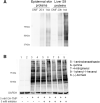
Immunoblot analysis of (A) BN rat liver S9 proteins and (B) epidermal skin proteins after 3 days of treatment with 400 mg/kg/day of TMP, 400 mg/kg/day of TMP=O (Ket1, Ket2), 100 mg/kg/day of TMP-OH (OH1, OH2) or 100 µl of 0.5% methylcellulose in PBS by gavage (C1−C3); (C) BN rat liver S9 proteins and (D) epidermal skin proteins from treatment with 0.2%–0.6% TMP in food for 5 weeks (TMP1−TMP4) or regular rodent meal (C1−C2). Numbers 1 − 4 denotes 4 different rats. Protein loading was 20 µg of liver protein and 40 µg of epidermal skin protein; primary antibody was diluted 1:5000 and 1:2000, respectively.
Figure 3.
Figure 3.
In vitro covalent binding of TMP and its metabolites to liver and epidermal skin proteins isolated from SD rats. Immunoblot analysis of (A) S9 liver or epidermal skin proteins incubated with TMP-S (SO3) or TMP-OH (OH), with a DMF solvent control; B, S9 liver proteins incubated with TMP, TMP=O, or TMP-OH. Protein loading was 25 µg, and the primary antibody was diluted 1:1000.
Figure 4.
Figure 4.
Immunoblot analysis of (A) epidermal skin or liver proteins incubated with 5 mM TMP-OH for 20 h or 1 week; B, freshly prepared S9 liver proteins incubated with various inhibitors (5 mM) for 30 min followed by incubation with 5 mM of TMP-OH for 20 h. Fifty micrograms of protein were loaded, and the primary antibody was diluted 1:1000.
Figure 5.
Figure 5.
Trimethoprim did not cause liver injury in female PD-1−/− mice co-treated with anti-CTLA-4 and 2% TMP in food. A, Serum ALT up to 3 weeks; B, serum GLDH up to 5 weeks. C, Immunoblot analysis of liver proteins from TMP-treated mice and BN rat (5 weeks). Protein loading was 20 μg, and primary antibody was diluted 1:5000. Values represent the mean ± SD. Analyzed for statistical significance by the unpaired t test ANOVA. p<0.05 was considered significant.
Figure 6.
Figure 6.
Analysis of the TMP-OH-sulfating activity of human SULTs. A, the results from a systematic analysis of all thirteen known human SULTs. B, the results from a concentration-dependent experiment using SULT1C4. The figure shows the autoradiographs taken from the TLC plates used for the analyses. In (A), lanes correspond to a control without enzyme and assay mixtures catalyzed by SULT1A1, SULT1A2, SULT1A3, SULT1B1, SULT1C2, SULT1C3, SULT1C4, SULT1E1, SULT2A1, SULT2B1a, SULT2B1b, SULT4A1, and SULT6B1, respectively. The final concentration of the substrate (TMP-OH) tested was 250 µM. The arrow indicates the position of [35S]sulfated TMP-OH. In (B), Lanes 1–3 correspond to control with substrate (TMP-OH; dissolved in DMSO) but without enzyme (SULT1C4), control without substrate and DMSO but with enzyme, and control without substrate but with DMSO and enzyme, respectively. Lanes 4 − 7 correspond to assay samples containing 50, 250, 500, and 1000 µM of TMP-OH, respectively.

References

    1. Bairam A. F., Rasool M. I., Alherz F. A., Abunnaja M. S., El Daibani A. A., Gohal S. A., Kurogi K., Sakakibara Y., Suiko M., Liu M. C. (2018). Sulfation of catecholamines and serotonin by SULT1A3 allozymes. Biochem. Pharmacol. 151, 104–113. - PMC - PubMed
    1. Baker C. A., Uno H., Johnson G. A. (1994). Minoxidil sulfation in the hair follicle. Skin Pharmacol. Physiol. 7, 335–339. - PubMed
    1. Brogden R. N., Carmine A. A., Heel R. C., Speight T. M., Avery G. S. (1982). Trimethoprim: A review of its antibacterial activity, pharmacokinetics and therapeutic use in urinary tract infections. Drugs 23, 405–430. - PubMed
    1. Fuchs E. (1983). Evolution and complexity of the genes encoding the keratins of human epidermal cells. J. Invest. Dermatol. 81, S141–S144. - PubMed
    1. Goldman J. L., Koen Y. M., Rogers S. A., Li K., Leeder J. S., Hanzlik R. P. (2016). Bioactivation of trimethoprim to protein-reactive metabolites in human liver microsomes. Drug Metab. Dispos. 44, 1603–1607. - PMC - PubMed

Publication types