

Chest X-ray in the emergency department during COVID-19 pandemic descending phase in Italy: correlation with patients' outcome
- PMID: 33394364
- PMCID: PMC7780606
- DOI: 10.1007/s11547-020-01327-3
Chest X-ray in the emergency department during COVID-19 pandemic descending phase in Italy: correlation with patients' outcome
Abstract
Purpose: The aims of our study are: (1) to estimate admission chest X-ray (CXR) accuracy during the descending phase of pandemic; (2) to identify specific CXR findings strictly associated with COVID-19 infection; and (3) to correlate lung involvement of admission CXR with patients' outcome.
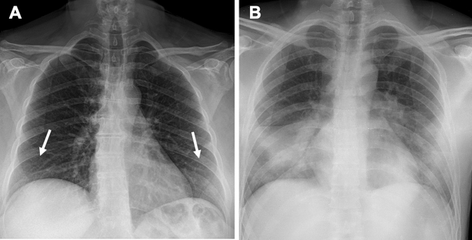
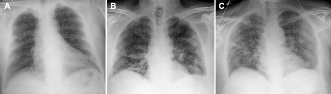
Materials and methods: We prospectively evaluated the admission CXR of 327 patients accessed to our institute during the Italian pandemic descending phase (April 2020). For each CXR were searched ground glass opacification (GGO), consolidation (CO), reticular-nodular opacities (RNO), nodules, excavations, pneumothorax, pleural effusion, vascular congestion and cardiac enlargement. For lung alterations was defined the predominance (upper or basal, focal or diffuse, central or peripheric, etc.). Then radiologists assessed whether CXRs were suggestive or not for COVID-19 infection. For COVID-19 patients, a prognostic score was applied and correlated with the patients' outcome.
Results: CXR showed 83% of specificity and 60% of sensitivity. GGO, CO, RNO and a peripheric, diffuse and basal prevalence showed good correlation with COVID-19 diagnosis. A logistic regression analysis pointed out GGO and a basal or diffuse distribution as independent predictors of COVID-19 diagnosis. The prognostic score showed good correlation with the patients' outcome.
Conclusion: In our study, admission CXR showed a fair specificity and a good correlation with patients' outcome. GGO and others CXR findings showed a good correlation with COVID-19 diagnosis; besides GGO a diffuse or bibasal distribution resulted in independent variables highly suggestive for COVID-19 infection thus enabling radiologists to signal to clinicians radiologically suspect patients during the pandemic descending phase.
Keywords: COVID-19 pneumonia; Chest radiograph; Diagnosis; Emergency department.
Conflict of interest statement
The authors declare that they have no conflict of interest related to the publication of this article.
Figures


References
-
- ACR Recommendations for the use of Chest Radiography and Computed Tomography (CT) for Suspected COVID-19 Infection (2020) American College of Radiology. https://www.acr.org/Advocacy-and-Economics/ACR-Position-Statements/Recom.... Accessed 22 Mar 2020
MeSH terms
LinkOut - more resources
Full Text Sources
Other Literature Sources
Medical
Miscellaneous
