Case Reports
. 2021 Dec;15(4):1308-1312.
doi: 10.1007/s12105-020-01264-7. Epub 2021 Jan 4.

Carcinoma Cuniculatum of the Larynx

Affiliations
Case Reports

Carcinoma Cuniculatum of the Larynx

Dario Marcotullio et al. Head Neck Pathol. 2021 Dec.

Abstract

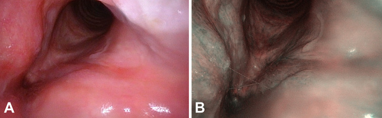
Carcinoma cuniculatum (CC) is a rare clinicopathologic variant of squamous cell carcinoma. Histologically, it is characterized by invasive growth of bland, acanthotic, and keratinizing squamous epithelium that forms multiple rabbit burrow-like, keratin-filled crypts and sinuses. We present a 51-year-old male smoker with CC of the left vocal cord. The tumor was staged T1a and the patient was disease-free 12 months after surgery. To our knowledge, this is the fourth case of CC of the larynx reported in the English literature and the first, due to its early diagnosis, where radical surgery was not performed. We highlight the necessity for awareness of this entity and coordination between otolaryngologists, radiologists, and pathologists for early diagnosis and organ-sparing surgical treatment.

Keywords: Carcinoma cuniculatum; Histological diagnosis; Larynx; Upper aero-digestive tract; Verrucous carcinoma; Vocal cords; Well-differentiated squamous cell carcinoma.


Conflict of interest statement

The authors declare that they have no conflict of interest.

Figures

Fig. 1
Fig. 1
Computed tomography images in the axial (a) and coronal (b) planes showing minimal local involvement of the left vocal cord with no extension to the close structures and no cervical lymphadenopathy
Fig. 2
Fig. 2
Low-power magnification of the lesion removed through microlaryngoscopy (a and b). The images were obtained from two different histological sections. The lesion consists of a branching, endophytic proliferation of keratinizing squamous epithelium forming crypts/sinuses that “burrow” within the superficial lamina propria. A florid inflammatory cell infiltrate is present in the lamina propria. Crypts/sinuses are lined by well-differentiated squamous epithelium and filled with keratin (c), that is focally intermixed with inflammatory cells (d). Dyskeratosis, intra-epithelial neutrophils, and micro-abscesses are illustrated in e. Staining: haematoxylin and eosin. Bars: 500 μm in a and b, 100 μm in c and 50 μm in d and e
Fig. 3
Fig. 3
A minimally branching, superficial crypt/sinus was observed in the sample obtained at cordectomy. The crypt/sinus is filled with keratin and associated with a florid inflammatory infiltrate within the lamina propria. Staining: haematoxylin and eosin. Bar: 150 μm
Fig. 4
Fig. 4
Flexible white-light (a) and NBI (b) laryngoscopy performed 6 months after surgery

References

    1. Aird I, Johnson HD, Lennox B, Stansfeld AG. Epithelioma cuniculatum: a variety of squamous carcinoma peculiar to the foot. Br J Surg. 1954;42:245–250. doi: 10.1002/bjs.18004217304. - DOI - PubMed
    1. Kao GF, Graham JH, Helwig EB. Carcinoma cuniculatum (verrucous carcinoma of the skin): a clinicopathologic study of 46 cases with ultrastructural observations. Cancer. 1982;49:2395–2403. doi: 10.1002/1097-0142(19820601)49:11<2395::aid-cncr2820491129>3.0.co;2-3. - DOI - PubMed
    1. Datar UV, Kale A, Mane D. Oral carcinoma cuniculatum: a new entity in the clinicopathological spectrum of oral squamous cell carcinoma. J Clin Diagn Res. 2017;11:37–39. doi: 10.7860/JCDR/2017/23437.9226. - DOI - PMC - PubMed
    1. Griffith CC, Goodstein LA, Tuluc M, Prestipino A, Cognetti D, Spiegel J, et al. A rare case of carcinoma cuniculatum involving the larynx in association with a saccular cyst. Head Neck Pathol. 2015;9:421–425. doi: 10.1007/s12105-014-0603-z. - DOI - PMC - PubMed
    1. Flieger S, Owinski T. Epithelioma cuniculatum an unusual form of mouth and jaw neoplasm. Czas Stomatol. 1977;30:395–401. - PubMed

Publication types

LinkOut - more resources