. 2021 Jan 4;19(1):e3001029.
doi: 10.1371/journal.pbio.3001029. eCollection 2021 Jan.

Cyclin-dependent Kinase 1 and Aurora Kinase choreograph mitotic storage and redistribution of a growth factor receptor

Affiliations

Cyclin-dependent Kinase 1 and Aurora Kinase choreograph mitotic storage and redistribution of a growth factor receptor

Christina D Cota et al. PLoS Biol. .

Abstract

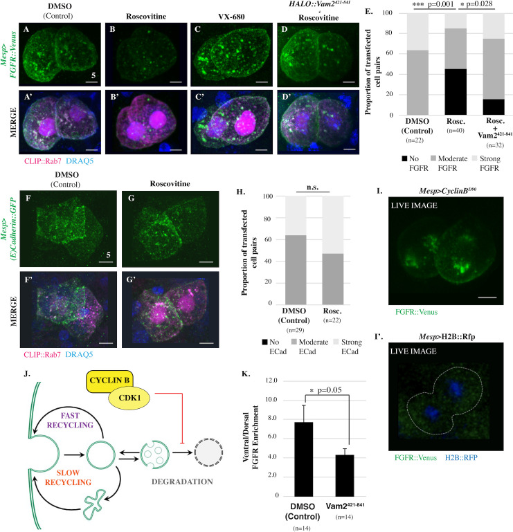
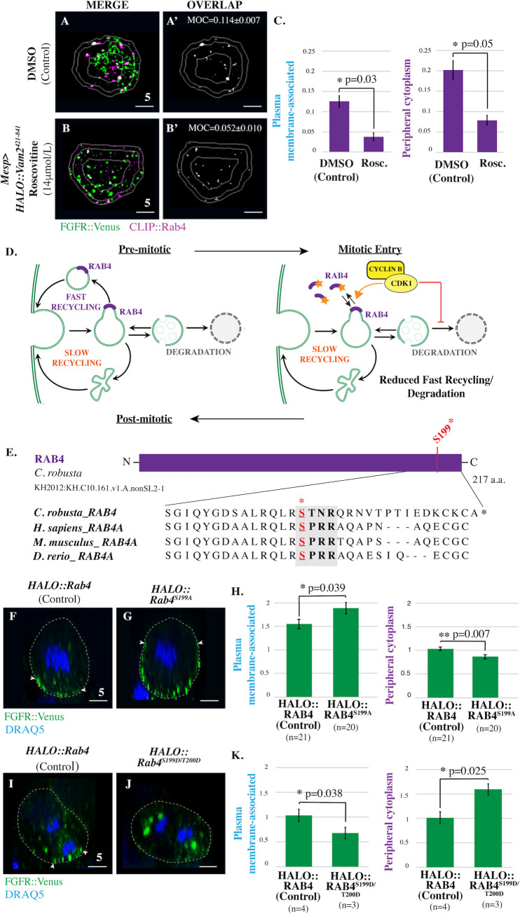
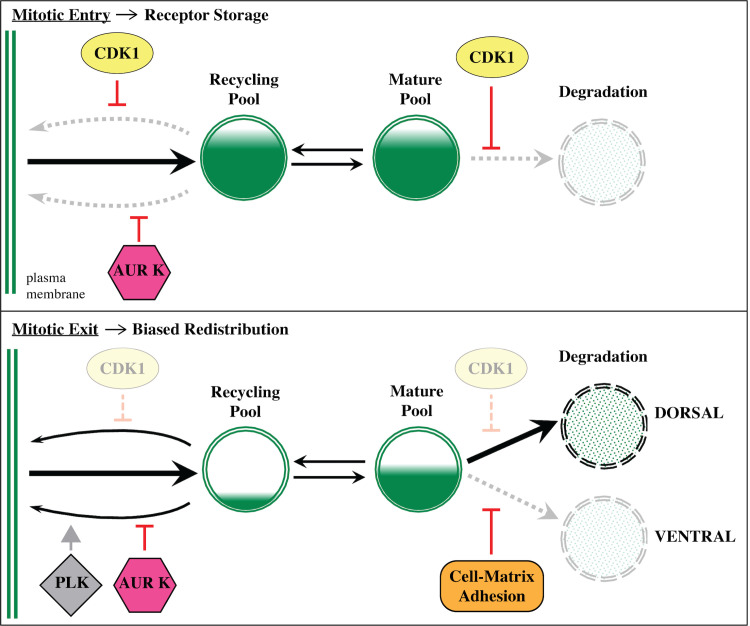
Endosomal trafficking of receptors and associated proteins plays a critical role in signal processing. Until recently, it was thought that trafficking was shut down during cell division. Thus, remarkably, the regulation of trafficking during division remains poorly characterized. Here we delineate the role of mitotic kinases in receptor trafficking during asymmetric division. Targeted perturbations reveal that Cyclin-dependent Kinase 1 (CDK1) and Aurora Kinase promote storage of Fibroblast Growth Factor Receptors (FGFRs) by suppressing endosomal degradation and recycling pathways. As cells progress through metaphase, loss of CDK1 activity permits differential degradation and targeted recycling of stored receptors, leading to asymmetric induction. Mitotic receptor storage, as delineated in this study, may facilitate rapid reestablishment of signaling competence in nascent daughter cells. However, mutations that limit or enhance the release of stored signaling components could alter daughter cell fate or behavior thereby promoting oncogenesis.


Conflict of interest statement

The authors have declared that no competing interests exist.

Figures

Fig 1
Fig 1. Mitotic trafficking of FGF receptors during founder cell division.
(A) Models depicting differential FGFR (green) redistribution during asymmetric founder cell division based on previous data (left panel) [20,41] along with a summary of endosomal pathways (right panel). For simplicity, schematics depict lateral views of a single founder cell. Regions of actin enrichment (purple; [40].) and adherent membrane (yellow, [41]) are indicated. (B-B”) Transverse sections and graphical summary depicting 3D-volumetric analysis of FGFR::VENUS distribution (quantified as regional enrichment; Methods) in a representative mitotic founder cell. Lines indicate region boundaries (white). Scale bars are indicated in micrometers. (C-D) Diagrammatic and graphical summaries of regional FGFR::VENUS enrichment (green) during founder cell division. Some regions are labeled with an a or b to denote that significant changes (p < 0.05) occurred within this region across cell cycle stages. Other regions are labeled n.s. to denote that no significant changes occurred for the indicated stages. Sample numbers for each stage are as follows: premitotic n = 50, prophase n = 36, metaphase n = 17, anaphase n = 24, and post-mitotic n = 34. Significance was determined using one-way ANOVA followed by Tukey multiple comparison test. Numerical values for all graphs can be found in S1 Data. ATM, Anterior Tail Muscle Cell; FGFR, Fibroblast Growth Factor Receptor; TVC, Trunk ventral cell/Cranial-cardiac progenitor.
Fig 2
Fig 2. Mitotic FGFR trafficking during founder cell division.
(A-C’) Masked/thresholded transverse sections and graphical summary depicting 3D-volumetric analysis of FGFR::VENUS/CLIP::RAB-GTPase colocalization (Manders’ overlap; MOC) in representative mitotic founder cells. Lines indicate region boundaries (white). Scale bars are indicated in micrometers. (D) Graphical summary of regional FGFR::VENUS enrichment (green) and FGFR::VENUS/ CLIP::RAB-GTPase colocalization (CLIP::RAB4, purple; CLIP::RAB7, orange; CLIP::RAB11, blue) during founder cell division. Some regions are labeled with an a or b to denote that significant changes (p < 0.05) occurred within this region across stages. Other regions are labeled n.s. to denote that no significant changes occurred for the indicated stages. (E-F’) Quantification of total (whole cell; E and F) and regional (E’-E”’, F’-F”’) FGFR::VENUS/ CLIP::RAB-GTPase colocalization during founder cell division showing significant changes in RAB7 and RAB11 values. Significance was determined using one-way ANOVA followed by Tukey multiple comparison test. Numerical values for all graphs can be found in S2 Data. (G) Model of mitotic FGFR trafficking illustrating stage-specific shifts as indicated. See also S2 Fig. FGFR, Fibroblast Growth Factor Receptor; MOC, Manders’ overlap coefficient.
Fig 3
Fig 3. CDK1 inhibits lysosomal degradation of FGFR.
(A-D’) Ventral projections of founder cell pairs electroporated with Mesp>FGFR::Venus alone or in combination with Mesp>HALO::Vam2421-841 as indicated and treated with vehicle (DMSO), Roscovitine (14 μmol/L) or VX-680 (21 μmol/L). In this experiment, Mesp>CLIP::Rab7 was included as a positive control for transfection. (E) Qualitative scoring of FGFR::VENUS intensity in transfected founder cell pairs. Significance was determined using Fisher exact test followed by Pearson chi-squared test. n = number of founder cell pairs scored. Treatment with AMG-900 (10 μmol/L) also had no significant impact on FGFR::VENUS intensity. Indeed, there was a nonsignificant increase in the number of cell pairs displaying strong FGFR::VENUS signal in the treated samples—45.8% ± 4.17 of AMG-900-treated cell pairs (n = 21) versus 34.5% ± 1.04 of DMSO-treated cell pairs (n = 42), p = 0.523. (F-G’) Ventral projections of founder cell pairs electroporated with Mesp>E-Cadherin::GFP and treated with vehicle (DMSO), or roscovitine (14 μmol/L). Mesp>CLIP::Rab7 was included as a positive control for transfection. (H) Qualitative scoring of E-CADHERIN::GFP intensity in transfected founder cell pairs. No significant differences found between treatments indicated. Significance was determined using Fisher exact test followed by Pearson chi-squared test. n = number of founder cell pairs scored. (I-I’) Ventral projection of FGFR::VENUS distribution in transgenic representative live founder cell pairs coelectroporated with Mesp>FGFR::Venus and Mesp>CyclinBΔ90 (I) or a control coelectroporated with Mesp>FGFR::Venus and Mesp>H2B::RFP (I’). Note that GFP/YFP signal in the heart founder lineage in the control (outlined by a white dashed line) are not above background levels. This image is representative of numerous observations of Mesp>FGFR::Venus in live embryos in which it is impossible to discern any signal leading to the standard use of antibody staining in fixed samples to assay FGFR localization. (J) Model depicting proposed CDK1-dependent inhibition of FGFR::VENUS degradation. (K) Quantification of FGFR::VENUS polarization in founder cells electroporated and treated as indicated. n = number of founder cells analyzed. Significance was determined using one-way ANOVA followed by Tukey multiple comparison test. Numerical values for all graphs can be found in S3 Data. Scale bars are indicated in micrometers. See also S3 Fig. CDK1, Cyclin-dependent Kinase 1; FGFR, Fibroblast Growth Factor Receptor; GFP, green fluorescent protein; YFP, yellow fluorescent protein.
Fig 4
Fig 4. CDK1 inhibits RAB4-dependent fast recycling of FGFR during mitotic entry.
(A-B’) Masked/thresholded transverse sections of founder cells electroporated with Mesp>FGFR::Venus alone or in combination with Mesp>HALO::Vam2421-841 and treated with vehicle (DMSO) or Roscovitine (14 μmol/L) as indicated. For clarity, images showing only the colocalized FGFR::VENUS/ CLIP::RAB-GTPase puncta in representative sections are provided (OVERLAP; Manders’ overlap; MOC) (A’ and B’). (C) Quantification of regional FGFR::VENUS/CLIP::RAB4 colocalization for founder cells electroporated and treated as indicated. (D) Model depicting proposed CDK1-dependent regulation of FGFR::VENUS trafficking. (E) Schematic depiction of C. robusta RAB4 protein. ClustalW alignment shows conservation of previously reported CDK1 phosphorylation motif (bold; [27]). Red asterisk indicates the serine residue phosphorylated by CDK1 in human cells. Putative phosphorylated serine residues in orthologs are indicated (S, red). (F-G) Lateral sections of prophase founder cells electroporated with either Mesp>FGFR::Venus along with either Mesp>HALO::Rab4 or Mesp>HALO::Rab4S199A as indicated. (H) Quantification of regional FGFR::VENUS enrichment in prophase founder cells electroporated as indicated. n = number of founder cells analyzed. (I-J) Lateral sections of anaphase founder cells electroporated with either Mesp>FGFR::Venus along with either Mesp>HALO::Rab4 or Mesp>HALO::Rab4S199D/T200D as indicated. (K) Quantification of regional FGFR::VENUS enrichment in anaphase founder cells electroporated as indicated. n = number of founder cells analyzed. Significance was determined using one-way ANOVA followed by Tukey multiple comparison test (C, H, K). Numerical values for all graphs can be found in S4 Data. Dashed lines indicate cell membranes that were delineated by phalloidin staining (F-G and I-J; red). White arrowheads (F-G and I-J) indicate dorsal boundaries of membrane-associated FGFR::VENUS puncta. Scale bars are indicated in micrometers. See also S4 Fig, S5 Fig and S6 Fig. CDK1, Cyclin-dependent Kinase 1; FGFR, Fibroblast Growth Factor Receptor; MOC, Manders’ overlap coefficient.
Fig 5
Fig 5. AurK promotes endosomal maturation and inhibits slow recycling of FGFR during mitotic entry.
(A-C) Lateral sections, graphical summary, and quantitative analysis of regional FGFR::VENUS enrichment for founder cells electroporated with Mesp>FGFR::Venus and treated with vehicle (DMSO) or VX-680 (21 μmol/L) as indicated. n = number of founder cells analyzed. (D-E) Masked/thresholded transverse sections of founder cells electroporated with Mesp>FGFR::Venus and Mesp>HALO::RAB11. For clarity, images showing only colocalized FGFR::VENUS/ CLIP::RAB-GTPase puncta in representative sections are provided (OVERLAP; Manders’ overlap; MOC) (D’ and E’). (F) Graphical summary of total (whole cell) or regional FGFR::VENUS/CLIP::RAB11 colocalization (Manders’ overlap). (G-H) Masked/thresholded transverse sections of founder cells electroporated with Mesp>FGFR::Venus and Mesp>HALO::RAB7. For clarity, images showing only colocalized FGFR::VENUS/ CLIP::RAB-GTPase puncta in representative sections are provided (MOC for panel H’ = 0.119±0.027) (G’ and H’). (I) Graphical summary of total (whole cell) or regional FGFR::VENUS/CLIP::RAB7 colocalization (Manders’ overlap). (J) Quantification of FGFR::VENUS ventral/dorsal polarization in founder cells treated with vehicle (DMSO) or VX-680 (21 μmol/L) as indicated. Significance was determined using one-way ANOVA followed by Tukey multiple comparison test (C, F, I, J). Numerical values for all graphs can be found in S5 Data. (K) Proposed model of CDK1 and AurK-dependent regulation of mitotic FGFR::VENUS trafficking during mitotic entry. In all micrographs, red dashed lines indicate cell membranes as delineated by phalloidin staining. Scale bars are indicated in micrometers. White arrowheads (A-B) indicate dorsal boundaries of membrane-associated FGFR::VENUS puncta. See also S7 Fig. AurK, Aurora Kinase; CDK1, Cyclin-dependent Kinase 1; FGFR, Fibroblast Growth Factor Receptor; MOC, Manders’ overlap coefficient.
Fig 6
Fig 6. Model for mitotic regulation of FGFR trafficking.
Diagrams illustrating hypothesized kinase-dependent shifts in trafficking and their impact on FGFR storage and redistribution during founder cell division. FGFR, Fibroblast Growth Factor Receptor.

References

    1. Boucrot E, Kirchhausen T. Endosomal recycling controls plasma membrane area during mitosis. Proc National Acad Sci. 2007;104:7939–44. 10.1073/pnas.0702511104 - DOI - PMC - PubMed
    1. Pellinen T, Tuomi S, Arjonen A, Wolf M, Edgren H, Meyer H, et al. Integrin Trafficking Regulated by Rab21 Is Necessary for Cytokinesis. Dev Cell. 2008;15:371–85. 10.1016/j.devcel.2008.08.001 - DOI - PubMed
    1. Wilson GM, Fielding AB, Simon GC, Yu X, Andrews PD, Hames RS, et al. The FIP3-Rab11 Protein Complex Regulates Recycling Endosome Targeting to the Cleavage Furrow during Late Cytokinesis. Mol Biol Cell. 2005;16:849–60. 10.1091/mbc.e04-10-0927 - DOI - PMC - PubMed
    1. Galvez T, Gilleron J, Zerial M, O’Sullivan GA. SnapShot: Mammalian Rab Proteins in Endocytic Trafficking. Cell. 2012;151:234–234.e2. 10.1016/j.cell.2012.09.013 - DOI - PubMed
    1. Stenmark H. Rab GTPases as coordinators of vesicle traffic. Nat Rev Mol Cell Biol. 2009;10:513–25. 10.1038/nrm2728 - DOI - PubMed

Publication types

MeSH terms