

Histological correlates of hippocampal magnetization transfer images in drug-resistant temporal lobe epilepsy patients
- PMID: 33395959
- PMCID: PMC7586233
- DOI: 10.1016/j.nicl.2020.102463
Histological correlates of hippocampal magnetization transfer images in drug-resistant temporal lobe epilepsy patients
Abstract
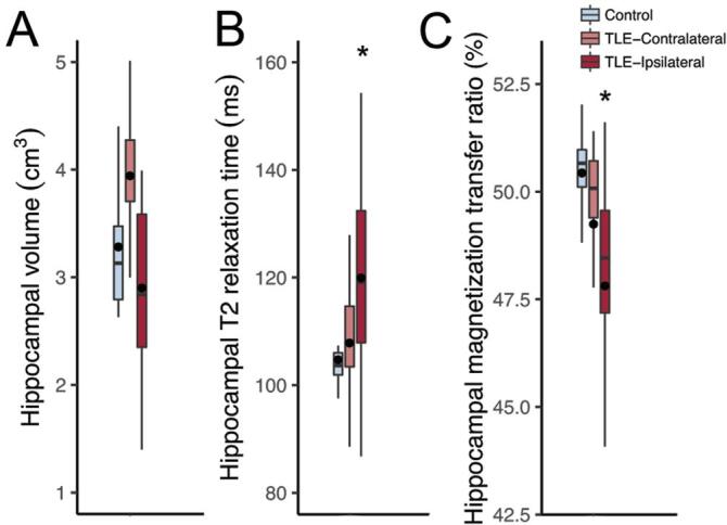
Objective: Temporal lobe epilepsy patients (TLE) often present with hippocampal atrophy, increased T2 relaxation, and reduced magnetization transfer ratio (MTR) in magnetic resonance images (MRI). The histological correlates of the reduced hippocampal MTR are so far unknown. Since MTR is dependent on the tissue's macromolecules, our aim was to evaluate the correlations between cellular populations, extracellular matrix molecules and the MTR in TLE patients.
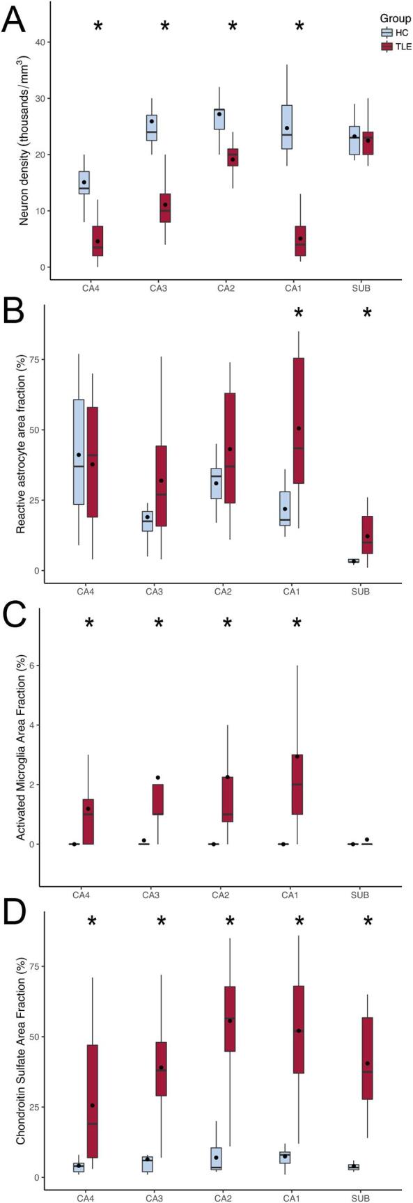
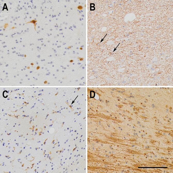
Methods: Patients with TLE (n = 26) and voluntaries (=20) were scanned in a 3 Tesla MRI scanner, and MTR images were calculated from 3DT1 sequences with magnetization pulse on resonance. Immunohistochemistry for neurons, reactive astrocytes, activated microglia, and extracellular matrix chondroitin sulfate were performed in formalin fixed, paraffin embedded tissues of TLE and autopsy controls (n = 10). Results were considered significant with adjusted p < 0.05.
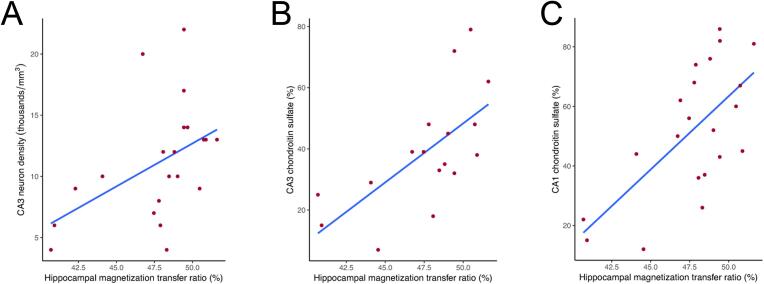
Results: Compared to the respective controls, TLE patients had reduced hippocampal MTR, increased reactive astrocytes and activated microglia, increased extracellular chondroitin sulfate, and reduced neuron density, compares to controls. MTR correlated positively with neuron density in CA3 and with chondroitin sulfate in CA3 and CA1. Multiple linear regressions reinforced the correlations between chondroitin sulfate and MTR.
Significance: Our data indicate that extracellular matrix molecules are the most significant histological correlates of magnetization transfer ratio in the hippocampus of TLE patients.
Keywords: Extracellular matrix; Hippocampal sclerosis; Magnetization transfer ratio; Neuron density; Temporal lobe epilepsy.
Copyright © 2020 The Authors. Published by Elsevier Inc. All rights reserved.
Figures
Similar articles
-
Individual hippocampal subfield assessment indicates that matrix macromolecules and gliosis are key elements for the increased T2 relaxation time seen in temporal lobe epilepsy.Epilepsia. 2017 Jan;58(1):149-159. doi: 10.1111/epi.13620. Epub 2016 Nov 18. Epilepsia. 2017. PMID: 27864825
-
Temporal lobe epilepsy patients with severe hippocampal neuron loss but normal hippocampal volume: Extracellular matrix molecules are important for the maintenance of hippocampal volume.Epilepsia. 2015 Oct;56(10):1562-70. doi: 10.1111/epi.13082. Epub 2015 Jul 27. Epilepsia. 2015. PMID: 26218733
-
T2 hyperintense signal in patients with temporal lobe epilepsy with MRI signs of hippocampal sclerosis and in patients with temporal lobe epilepsy with normal MRI.Epilepsy Behav. 2015 May;46:103-8. doi: 10.1016/j.yebeh.2015.04.001. Epub 2015 May 1. Epilepsy Behav. 2015. PMID: 25936278
-
MRI morphology of the hippocampus in drug-resistant temporal lobe epilepsy: Shape inflation of left hippocampus and correlation of right-sided hippocampal volume and shape with visuospatial function in patients with right-sided TLE.J Clin Neurosci. 2019 Sep;67:68-74. doi: 10.1016/j.jocn.2019.06.019. Epub 2019 Jun 17. J Clin Neurosci. 2019. PMID: 31221579
-
MRI-negative temporal lobe epilepsy-What do we know?Epilepsia. 2017 May;58(5):727-742. doi: 10.1111/epi.13699. Epub 2017 Mar 7. Epilepsia. 2017. PMID: 28266710 Review.
Cited by
-
Contribution of perineuronal nets to hyperexcitability in pilocarpine-induced status epilepticus.Epilepsia. 2025 Jun 28:10.1111/epi.18489. doi: 10.1111/epi.18489. Online ahead of print. Epilepsia. 2025. PMID: 40580058
-
Repurposing dimethyl fumarate as an antiepileptogenic and disease-modifying treatment for drug-resistant epilepsy.J Transl Med. 2023 Nov 8;21(1):796. doi: 10.1186/s12967-023-04695-2. J Transl Med. 2023. PMID: 37940957 Free PMC article.
References
-
- Blumcke I., Spreafico R., Haaker G., Coras R., Kobow K., Bien C.G. Histopathological Findings in Brain Tissue Obtained during Epilepsy Surgery. The New England J. Med. 2017;377:1648–1656. - PubMed
-
- Blumcke I., Thom M., Aronica E., Armstrong D.D., Bartolomei F., Bernasconi A. International consensus classification of hippocampal sclerosis in temporal lobe epilepsy: a Task Force report from the ILAE Commission on Diagnostic Methods. Epilepsia. 2013;54:1315–1329. - PubMed
-
- Crespel A., Coubes P., Rousset M.C., Brana C., Rougier A., Rondouin G. Inflammatory reactions in human medial temporal lobe epilepsy with hippocampal sclerosis. Brain Res. 2002;952:159–169. - PubMed
-
- Van Paesschen W., Revesz T., Duncan J.S., King M.D., Connelly A. Quantitative neuropathology and quantitative magnetic resonance imaging of the hippocampus in temporal lobe epilepsy Annals of neurology. 1997;42:756–766. - PubMed
-
- Perosa S.R., Porcionatto M.A., Cukiert A., Martins J.R., Passeroti C.C., Amado D. Glycosaminoglycan levels and proteoglycan expression are altered in the hippocampus of patients with mesial temporal lobe epilepsy. Brain Res. Bull. 2002;58:509–516. - PubMed
Publication types
MeSH terms
Substances
LinkOut - more resources
Full Text Sources
Medical
Miscellaneous
