

Decreased grey matter in the postural control network is associated with lateral flexion of the trunk in Parkinson's disease
- PMID: 33395964
- PMCID: PMC7645287
- DOI: 10.1016/j.nicl.2020.102469
Decreased grey matter in the postural control network is associated with lateral flexion of the trunk in Parkinson's disease
Abstract
Background: Disruption of central networks, particularly of those responsible for integrating multimodal afferents in a spatial reference frame, were proposed in the pathophysiology of lateral trunk flexion in Parkinson's disease (PD). Knowledge about the underlying neuroanatomical structures is limited.
Objective: To investigate if decreased focal grey matter (GM) is associated with trunk flexion to the side and if the revealed GM clusters correlate with a disturbed perception of verticality in PD.
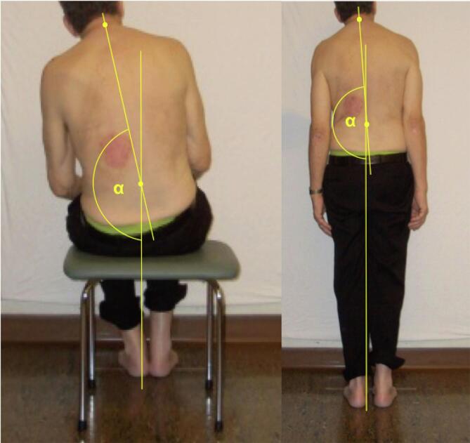
Methods: 37 PD patients with and without lateral trunk flexion were recruited. Standardized photos were taken from each patient and trunk orientation was measured by a blinded rater. Voxel-based morphometry (VBM) was used to detect associated clusters of decreased GM. The subjective visual vertical (SVV) was assessed as a marker for perception of verticality and SVV estimates were correlated with GM clusters.
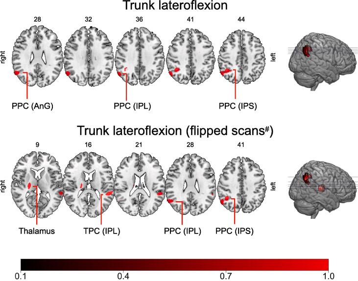
Results: VBM revealed clusters of decreased GM in the right posterior parietal cortex and in the right thalamus were associated with lateral trunk flexion. The SVV correlated with the extent of trunk flexion, and the side of the SVV tilt correlated with the side of trunk flexion. GM values from the thalamus correlated with the SVV estimates.
Conclusions: We report an association between neurodegenerative changes within the posterior parietal cortex and the thalamus and lateral trunk flexion in PD. These brain structures are part of a network proposed to be engaged in postural control and spatial self-perception. Disturbed perception of verticality points to a shifted egocentric spatial reference as an important pathophysiological feature.
Keywords: Lateral trunk flexion; MRI; Parkinson’s disease; Pisa syndrome; Subjective visual vertical.
Copyright © 2020 The Authors. Published by Elsevier Inc. All rights reserved.
Conflict of interest statement
The authors declare that they have no known competing financial interests or personal relationships that could have appeared to influence the work reported in this paper.
F. Brugger: Stock Ownership in medically-related fields: None; Intellectual Property Rights: None; Consultancies: None; Expert Testimony: None; Advisory Boards: None; Employment: Kantonsspital St. Gallen, Switzerland; Partnerships: None; Contracts: None; Honoraria: Speaker’s honorarium from Zambon Switzerland; Royalties: Südwestdeutscher Verlag für Hochschulschriften; Grants: Baasch-Medicus stipend, research grant by the KSSG Forschungskommission, travel grant by Abbvie Switzerland; Other: None.
Conflict of interests: none
J. Walch: Stock Ownership in medically-related fields: None; Intellectual Property Rights: None; Consultancies: None; Expert Testimony: None; Advisory Boards: None; Employment: Kantonsspital St. Gallen, Switzerland; Partnerships: None; Contracts: None; Honoraria: None; Royalties: None; Grants: Travel grant by Abbvie Switzerland; Other: None.
Conflict of interests: none
S. Hägele-Link: Stock Ownership in medically-related fields: None; Intellectual Property Rights: None; Consultancies: None; Expert Testimony: None; Advisory Boards: none; Employment: Kantonsspital St. Gallen, Switzerland; Partnerships: None; Contracts: None; Honoraria: Speaker’s honorarium from Abbvie Switzerland; Royalties: None; Grants: None; Other: None.
Conflict of interests: none
E. Abela: Stock Ownership in medically-related fields: None; Intellectual Property Rights: None; Consultancies: None; Expert Testimony: None; Advisory Boards: None; Employment: Inselspital Berne; Partnerships: None; Contracts: None; Honoraria: None; Royalties: None; Grants: SNF 33CM30-124089, and NIHR BRC Preparatory Fellowship from King’s College London; Other: None. Conflict of interests: none
M. Galovic: Stock Ownership in medically-related fields: None; Intellectual Property Rights: None; Consultancies: None; Expert Testimony: None; Advisory Boards: None; Employment: University Hospital Zurich, Switzerland; Partnerships: None; Contracts: None; Honoraria: None; Royalties: None; Grants: Epilepsy Research UK; Other: None.
Conflict of interests: none
G. Kägi: Stock Ownership in medically-related fields: None; Intellectual Property Rights: None; Consultancies: None; Expert Testimony: None; Advisory Boards: Bayer, Zambon; Employment: Kantonsspital St. Gallen, Switzerland; Partnerships: None; Contracts: None; Honoraria: none; Royalties: None; Grants: Swiss Parkinson Association, Swiss Heart Foundation; Other: None.
Figures
Similar articles
-
Cortical involvement of lateral trunk flexion and verticality misperception in Parkinson's disease.Brain Commun. 2025 Jan 27;7(1):fcaf040. doi: 10.1093/braincomms/fcaf040. eCollection 2025. Brain Commun. 2025. PMID: 39926614 Free PMC article.
-
Disturbance of verticality perception and postural dysfunction in Parkinson's disease.Acta Neurol Scand. 2018 Feb;137(2):212-217. doi: 10.1111/ane.12859. Epub 2017 Oct 23. Acta Neurol Scand. 2018. PMID: 29063605
-
Haptic perception of verticality correlates with postural and balance deficits in patients with Parkinson's disease.Parkinsonism Relat Disord. 2019 Sep;66:45-50. doi: 10.1016/j.parkreldis.2019.06.026. Epub 2019 Jul 2. Parkinsonism Relat Disord. 2019. PMID: 31279636
-
Voxelwise meta-analysis of gray matter anomalies in Parkinson variant of multiple system atrophy and Parkinson's disease using anatomic likelihood estimation.Neurosci Lett. 2015 Feb 5;587:79-86. doi: 10.1016/j.neulet.2014.12.007. Epub 2014 Dec 5. Neurosci Lett. 2015. PMID: 25484255
-
Lateropulsion with active pushing in stroke patients: its link with lesion location and the perception of verticality. A systematic review.Top Stroke Rehabil. 2023 Apr;30(3):281-297. doi: 10.1080/10749357.2022.2026563. Epub 2022 Feb 1. Top Stroke Rehabil. 2023. PMID: 35102816
Cited by
-
[Clinical application progress of subjective visual vertical test].Lin Chuang Er Bi Yan Hou Tou Jing Wai Ke Za Zhi. 2022 Nov;36(11):884-887;892. doi: 10.13201/j.issn.2096-7993.2022.11.016. Lin Chuang Er Bi Yan Hou Tou Jing Wai Ke Za Zhi. 2022. PMID: 36347586 Free PMC article. Review. Chinese.
-
Therapeutic Intervention for Trunk Control Impairments in Central Nervous System Disorders: A Comprehensive Review of Methods and Efficacy.Prog Rehabil Med. 2025 Jan 16;10:20250002. doi: 10.2490/prm.20250002. eCollection 2025. Prog Rehabil Med. 2025. PMID: 39822314 Free PMC article. Review.
-
Factors Contributing to the Severity and Laterality of Pisa Syndrome in Parkinson's Disease.Front Aging Neurosci. 2022 Jan 3;13:716990. doi: 10.3389/fnagi.2021.716990. eCollection 2021. Front Aging Neurosci. 2022. PMID: 35046790 Free PMC article.
References
-
- Baier B., Janzen J., Muller-Forell W., Fechir M., Muller N., Dieterich M. Pusher syndrome: its cortical correlate. J. Neurol. 2012;259(2):277–283. - PubMed
-
- Baier B., Suchan J., Karnath H.O., Dieterich M. Neural correlates of disturbed perception of verticality. Neurology. 2012;78(10):728–735. - PubMed
-
- Baier B., Thomke F., Wilting J., Heinze C., Geber C., Dieterich M. A pathway in the brainstem for roll-tilt of the subjective visual vertical: evidence from a lesion-behavior mapping study. The Journal of neuroscience : the official journal of the Society for Neuroscience. 2012;32(43):14854–14858. - PMC - PubMed
-
- Baier B., Conrad J., Stephan T., Kirsch V., Vogt T., Wilting J., Müller-Forell W., Dieterich M. Vestibular thalamus: Two distinct graviceptive pathways. Neurology. 2016;86(2):134–140. - PubMed
-
- Barra J., Marquer A., Joassin R., Reymond C., Metge L., Chauvineau V., Perennou D. Humans use internal models to construct and update a sense of verticality. Brain : a journal of neurology. 2010;133(Pt 12):3552–3563. - PubMed
MeSH terms
LinkOut - more resources
Full Text Sources
Medical
