

Heart development in two populations of Atlantic killifish (Fundulus heteroclitus) following exposure to a polycyclic aromatic hydrocarbon mixture
- PMID: 33396103
- PMCID: PMC7837385
- DOI: 10.1016/j.ecoenv.2020.111580
Heart development in two populations of Atlantic killifish (Fundulus heteroclitus) following exposure to a polycyclic aromatic hydrocarbon mixture
Abstract
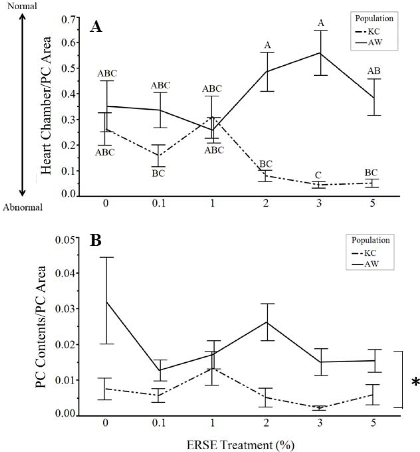
Historic industrial pollution of the Elizabeth River, Virginia resulted in polycyclic aromatic hydrocarbon (PAH) contamination in sediments. Atlantic killifish (Fundulus heteroclitus) inhabiting the Atlantic Wood (AW) industrial site adapted to complex PAH mixture at this Superfund site. Their embryos have proved highly resistant to cardiac abnormalities indicative of PAH toxicity. In this study, embryos spawned from adults collected at AW and King's Creek (KC), a reference site, were exposed at 24 h post fertilization (hpf) to Elizabeth River Sediment Extract (ERSE), a complex PAH mixture, in a range of concentrations (0, 5.04, 50.45, 100.90, 151.35, or 252.25 µg/L total PAHs). Embryos were processed for histology at 144 hpf to enable evaluations of hearts at tissue and cellular levels. Morphometry and severity scoring were used to evaluate the extent of alterations. Unexposed embryos were similar in both populations. ERSE exposure resulted in multiple changes to hearts of KC embryos but not AW. Alterations were particularly evident in KC embryos exposed to concentrations above 1% ERSE (50.45 µg/L), which had thinner ventricular walls and larger pericardial edema. Individuals with moderate pericardial edema maintained arrangement and proximity of heart chambers, but changes were seen in ventricular myocytes. Severe pericardial edema was prevalent in exposed KC embryos and typically resulted in tube heart formation. Ventricles of tube hearts had very thin walls composed of small, basophilic cells and lacked trabeculae. Edematous pericardial fluid contained small amounts of proteinaceous material, as did controls, and was free of cells. This fluid was primarily unstained, suggesting water influx due to increased permeability. The use of histological approaches provided more specific detail for tissue and cellular effects in hearts of embryos exposed to PAHs and enabled understanding of potential links to later life effects of early life exposure.
Keywords: Fundulus; Heart development; Histology; Morphometry; PAHs.
Copyright © 2020 The Authors. Published by Elsevier Inc. All rights reserved.
Conflict of interest statement
Authors have no competing interests.
Declaration of interests
The authors declare that they have no known competing financial interests or personal relationships that could have appeared to influence the work reported in this paper.
Figures


Similar articles
-
Later life swimming performance and persistent heart damage following subteratogenic PAH mixture exposure in the Atlantic killifish (Fundulus heteroclitus).Environ Toxicol Chem. 2017 Dec;36(12):3246-3253. doi: 10.1002/etc.3877. Epub 2017 Aug 24. Environ Toxicol Chem. 2017. PMID: 28585726 Free PMC article.
-
Hepatic Responses of Juvenile Fundulus heteroclitus from Pollution-adapted and Nonadapted Populations Exposed to Elizabeth River Sediment Extract.Toxicol Pathol. 2016 Jul;44(5):738-48. doi: 10.1177/0192623316636717. Epub 2016 Mar 18. Toxicol Pathol. 2016. PMID: 26992886 Free PMC article.
-
Compound- and mixture-specific differences in resistance to polycyclic aromatic hydrocarbons and PCB-126 among Fundulus heteroclitus subpopulations throughout the Elizabeth River estuary (Virginia, USA).Environ Sci Technol. 2013 Sep 17;47(18):10556-66. doi: 10.1021/es401604b. Epub 2013 Sep 5. Environ Sci Technol. 2013. PMID: 24003986 Free PMC article.
-
Characterization of the recalcitrant CYP1 phenotype found in Atlantic killifish (Fundulus heteroclitus) inhabiting a Superfund site on the Elizabeth River, VA.Aquat Toxicol. 2010 Aug 1;99(1):33-41. doi: 10.1016/j.aquatox.2010.03.015. Epub 2010 Apr 14. Aquat Toxicol. 2010. PMID: 20471113 Free PMC article.
-
The Elizabeth River Story: A Case Study in Evolutionary Toxicology.J Toxicol Environ Health B Crit Rev. 2015;18(6):259-98. doi: 10.1080/15320383.2015.1074841. Epub 2015 Oct 27. J Toxicol Environ Health B Crit Rev. 2015. PMID: 26505693 Free PMC article. Review.
Cited by
-
Transcriptomic and Methylomic Analyses Show Significant Shifts in Biosynthetic Processes and Reduced Intrapopulation Gene Expression Variance in PAH-Adapted Atlantic Killifish.Environ Sci Technol. 2024 Nov 26;58(47):20859-20872. doi: 10.1021/acs.est.4c06845. Epub 2024 Nov 17. Environ Sci Technol. 2024. PMID: 39552013
References
-
- Annunziato K, Cooper KR, The Impact of Early Developmental Exposure to Stressors Related to Individual Fitness in Aquatic Organisms and the Subsequent Reproductive Success and Failure on Populations In: Burggren W, Dubansky B, Eds.), Development and Environment. Springer International Publishing, Cham, 2018, pp. 115–153.
-
- Armstrong PB, Child JS, 1965. Stages in the normal development of Fundulus heteroclitus. Biological Bulletin. 128, 143–168. 10.2307/1539545. - DOI
MeSH terms
Substances
Grants and funding
LinkOut - more resources
Full Text Sources
Other Literature Sources
