Review
. 2020 Dec 31;13(1):47.
doi: 10.3390/pharmaceutics13010047.

Optimizations of In Vitro Mucus and Cell Culture Models to Better Predict In Vivo Gene Transfer in Pathological Lung Respiratory Airways: Cystic Fibrosis as an Example

Affiliations
Review

Optimizations of In Vitro Mucus and Cell Culture Models to Better Predict In Vivo Gene Transfer in Pathological Lung Respiratory Airways: Cystic Fibrosis as an Example

Rosy Ghanem et al. Pharmaceutics. .

Abstract

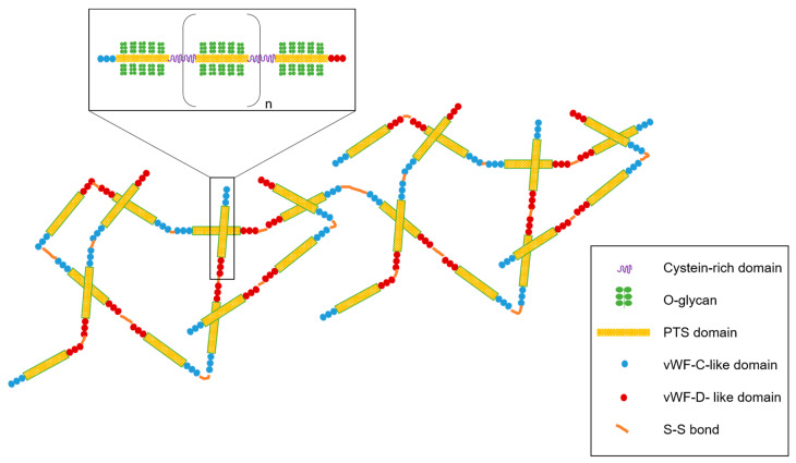
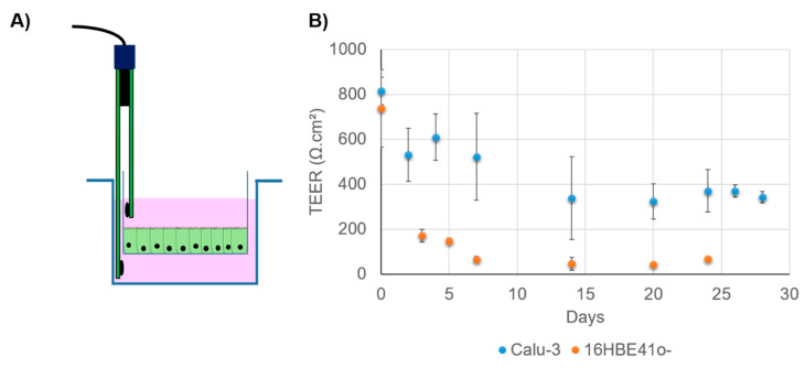
The respiratory epithelium can be affected by many diseases that could be treated using aerosol gene therapy. Among these, cystic fibrosis (CF) is a lethal inherited disease characterized by airways complications, which determine the life expectancy and the effectiveness of aerosolized treatments. Beside evaluations performed under in vivo settings, cell culture models mimicking in vivo pathophysiological conditions can provide complementary insights into the potential of gene transfer strategies. Such models must consider multiple parameters, following the rationale that proper gene transfer evaluations depend on whether they are performed under experimental conditions close to pathophysiological settings. In addition, the mucus layer, which covers the epithelial cells, constitutes a physical barrier for gene delivery, especially in diseases such as CF. Artificial mucus models featuring physical and biological properties similar to CF mucus allow determining the ability of gene transfer systems to effectively reach the underlying epithelium. In this review, we describe mucus and cellular models relevant for CF aerosol gene therapy, with a particular emphasis on mucus rheology. We strongly believe that combining multiple pathophysiological features in single complex cell culture models could help bridge the gaps between in vitro and in vivo settings, as well as viral and non-viral gene delivery strategies.

Keywords: airway epithelium; cystic fibrosis; gene delivery; in vitro model; mucus.


Conflict of interest statement

The authors declare no conflict of interest.

Figures

Figure 1
Figure 1
Schematic representation of the respiratory system and the use of an aerosol to target the pulmonary epithelium. (A) The upper and lower respiratory tract with an emphasis on a bronchial section constituted by mucous glands and smooth muscles. In the case of cystic fibrosis (CF), sticky mucus is overproduced and creates a bronchial plug. (B) Pseudostratified airway epithelium composed of goblet, ciliated, and basal cells. The mucosa is covered with airways surface liquid (ASL) which contains two layers, a periciliary liquid layer directly in contact with the cilia and mucus layer. CF mucus is colonized with opportunistic bacteria such as Staphylococcus aureus and Pseudomonas aeruginosa. These bacteria form a biofilm participating in the viscous and dense aspect of the CF mucus. When nanoparticles (NPs) such as lipoplexes are aerosolized, they have to cross the mucus barrier before reaching the epithelial cells to deliver therapeutic compounds such as acids nucleic construction.
Figure 2
Figure 2
General structure of mucins, the main constituent of mucus. Mucin contains the PTS (proline, threonine, and serine) domain and supporting O-glycan. The PTS domain is interspaced with cysteine-rich domains. Mucin glycopolymer ends with the von Willebrand factor (vWF) domain in N- and C-terminal regions. Mucin polymerized via disulfide bonds and mucin polymers interact with each other via hydrophobic and electrostatic interactions.
Figure 3
Figure 3
The pulmonary epithelium is maintained by adherent junctions and the tight junction (TJ) complex composed of occludin, claudins, and junction adhesion molecules (JAM). Zona occludens (ZO) is directly attached to actin and connects the TJ to the cytoskeleton, notably actin F.
Figure 4
Figure 4
(A) Scanning electron microscopy of 16HBE41o- and Calu-3 cultivated in the liquid–liquid interphase (LLI) or air–liquid interphase (ALI) for 3 weeks after seeding. When cultivated under ALI conditions, unlike 16HBE41o-, Calu-3 cells produce mucus and form microvilli. For all pictures, the scale bar corresponds to 20.0 µm. (B) Transition from LLI culture to ALI allowing cell differentiation and in some case, depending on the cell line, mucus production.
Figure 5
Figure 5
(A) Transepithelial electrical resistance (TEER) measurement using a Transwell and an electrode. (B) Evolution of TEER in Calu-3 and 16HBE41o- when cultivated in ALI conditions. Cells are seeded in a Transwell (150,000 cells per insert) and cultivated in LLI conditions for one week. Then, the apical media are removed. TEER is monitored for 25 to 30 days. Data are averages of at least three Transwells and are expressed in Ω·cm2. Calu-3 exhibits a higher TEER value, demonstrating its ability to form TJ under this condition.

References

    1. Kim N., Duncan G.A., Hanes J., Suk J.S. Barriers to inhaled gene therapy of obstructive lung diseases: A review. J. Control. Release. 2016;240:465. doi: 10.1016/j.jconrel.2016.05.031. - DOI - PMC - PubMed
    1. O’Sullivan B.P., Freedman S.D. Cystic fibrosis. Lancet. 2009;373:1891–1904. doi: 10.1016/S0140-6736(09)60327-5. - DOI - PubMed
    1. Ratjen F., Rietschel E., Griese M., Ballmann M., Kleinau I., Doring G., Reinhardt D., Paul K. Fractional analysis of bronchoalveolar lavage fluid cytology in cystic fibrosis patients with normal lung function. Bronchoalveolar lavage for the evaluation of anti-inflammatory treatment (BEAT) study group. Eur. Respir. J. 2000;15:141–145. doi: 10.1183/09031936.00.15114100. - DOI - PubMed
    1. Reinhardt N., Chen C.I.U., Loppow D., Schink T., Kleinau I., Jörres R.A., Wahn U., Magnussen H., Paul K.P. Cellular profiles of induced sputum in children with stable cystic fibrosis: Comparison with BAL. Eur. Respir. J. 2003;22:497–502. doi: 10.1183/09031936.03.00043603. - DOI - PubMed
    1. Jackson A.D. Airway goblet-cell mucus secretion. Trends Pharmacol. Sci. 2001;22:39–45. doi: 10.1016/S0165-6147(00)01600-X. - DOI - PubMed