Review
. 2020 Dec 31;10(1):51.
doi: 10.3390/cells10010051.

Role of Cardiac Macrophages on Cardiac Inflammation, Fibrosis and Tissue Repair

Affiliations
Review

Role of Cardiac Macrophages on Cardiac Inflammation, Fibrosis and Tissue Repair

William P Lafuse et al. Cells. .

Abstract

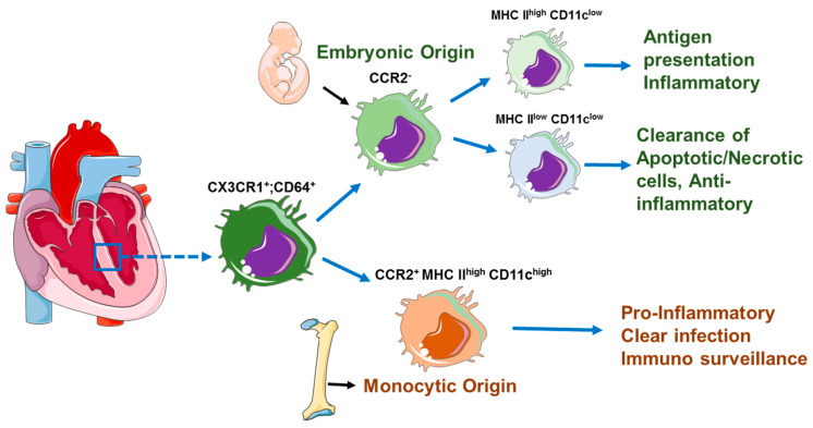
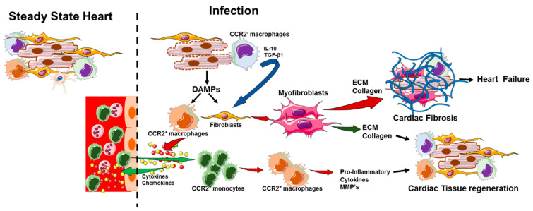
The immune system plays a pivotal role in the initiation, development and resolution of inflammation following insult or damage to organs. The heart is a vital organ which supplies nutrients and oxygen to all parts of the body. Heart failure (HF) has been conventionally described as a disease associated with cardiac tissue damage caused by systemic inflammation, arrhythmia and conduction defects. Cardiac inflammation and subsequent tissue damage is orchestrated by the infiltration and activation of various immune cells including neutrophils, monocytes, macrophages, eosinophils, mast cells, natural killer cells, and T and B cells into the myocardium. After tissue injury, monocytes and tissue-resident macrophages undergo marked phenotypic and functional changes, and function as key regulators of tissue repair, regeneration and fibrosis. Disturbance in resident macrophage functions such as uncontrolled production of inflammatory cytokines, growth factors and inefficient generation of an anti-inflammatory response or unsuccessful communication between macrophages and epithelial and endothelial cells and fibroblasts can lead to aberrant repair, persistent injury, and HF. Therefore, in this review, we discuss the role of cardiac macrophages on cardiac inflammation, tissue repair, regeneration and fibrosis.

Keywords: cardiac inflammation; cardiac macrophages; fibrosis; tissue repair.


Conflict of interest statement

The authors declare no conflict of interest.

Figures

Figure 1
Figure 1
Cellular components of steady-state heart. Heart tissue consists of endothelial cells, myocytes, fibroblasts, pericytes, mesenchymal cells, and various types of immune cells. The macrophages are the major immune cell population in the resting heart and are found in the interstitium and around endothelial cells. Inflammatory monocytes and neutrophils are not present in myocardial tissue but can be observed upon tissue damage. Mast cells, dendritic cells, B cells, NK cells and regulatory T cells are found sporadically in cardiac tissue.
Figure 2
Figure 2
Function of CCR2+ and CCR2 cardiac-resident macrophages in the resting heart. An adult heart contains CX3CR1+, CD64+, CCR2+ and CCR2-resident macrophages. The CCR2-resident macrophages enter the heart during embryonic development and possess the capability to self-proliferate. These macrophages are further divided into two groups based on MHC II expression. The CCR2 MHC IIhigh macrophages perform antigen presentation, whereas the CCR2 MHC IIlow CD11clow macrophage plays an important role in clearance of apoptotic cells and express anti-inflammatory mediators. The CCR2+ MHC IIhigh CD11chigh originate from bone marrow progenitors and perform immune surveillance functions and protect the heart from infection.
Figure 3
Figure 3
Cardiac immune response during infection and tissue repair. During early infection, cardiomyocytes undergo necrosis and release endogenous danger-associated molecular patterns (DAMPs), which activate resident mast cells and CCR2+ macrophages to release pro-inflammatory cytokines, such as TNFα and IL-1β, and chemokines, which activate endothelial cell dilation and recruit activated CCR2+ monocytes and neutrophils. The CCR2+ monocytes differentiate into macrophages, which together with neutrophils create an inflammatory environment and clear the invaded pathogens. IL-10 and TGFβ1 are released by the action of DAMPs directly on fibroblasts or from CCR2-macrophages that have efferocytosed necrotic cardiomycytes. IL-10 and TGF-β1 then activates fibroblasts to transdifferentiate into myofibroblasts. The myofibroblasts produce extracellular matrix (ECM) and collagen, fill in the gap, and regenerate cardiac tissue. However, increased cardiomyocyte death and cardiac inflammation leads to enhanced myofibroblast activation and uncontrolled production of ECM and collagen which accumulate in the injured heart tissue and cause cardiac fibrosis.

References

    1. Gordon S., Martinez-Pomares L. Physiological roles of macrophages. Pflugers Arch. 2017;469:365–374. doi: 10.1007/s00424-017-1945-7. - DOI - PMC - PubMed
    1. Roth G.A., Johnson C., Abajobir A., Abd-Allah F., Abera S.F., Abyu G., Ahmed M., Aksut B., Alam T., Alam K., et al. Global, Regional, and National Burden of Cardiovascular Diseases for 10 Causes, 1990 to 2015. J. Am. Coll. Cardiol. 2017;70:1–25. doi: 10.1016/j.jacc.2017.04.052. - DOI - PMC - PubMed
    1. Benjamin E.J., Muntner P., Alonso A., Bittencourt M.S., Callaway C.W., Carson A.P., Chamberlain A.M., Chang A.R., Cheng S., Das S.R., et al. Heart Disease and Stroke Statistics-2019 Update: A Report from the American Heart Association. Circulation. 2019;139:e56–e528. doi: 10.1161/CIR.0000000000000659. - DOI - PubMed
    1. Frangogiannis N.G. Cardiac fibrosis: Cell biological mechanisms, molecular pathways and therapeutic opportunities. Mol. Asp. Med. 2019;65:70–99. doi: 10.1016/j.mam.2018.07.001. - DOI - PubMed
    1. Hulsmans M., Sam F., Nahrendorf M. Monocyte and macrophage contributions to cardiac remodeling. J. Mol. Cell. Cardiol. 2016;93:149–155. doi: 10.1016/j.yjmcc.2015.11.015. - DOI - PMC - PubMed

Publication types

MeSH terms

Substances