. 2020 Dec 31;22(1):372.
doi: 10.3390/ijms22010372.

Polycyclic Aromatic Hydrocarbons Activate the Aryl Hydrocarbon Receptor and the Constitutive Androstane Receptor to Regulate Xenobiotic Metabolism in Human Liver Cells

Affiliations

Polycyclic Aromatic Hydrocarbons Activate the Aryl Hydrocarbon Receptor and the Constitutive Androstane Receptor to Regulate Xenobiotic Metabolism in Human Liver Cells

Lisa Goedtke et al. Int J Mol Sci. .

Abstract

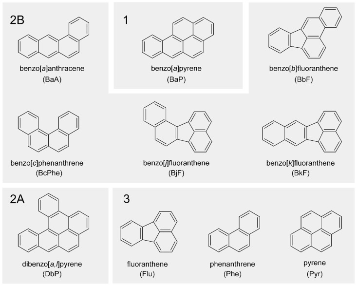
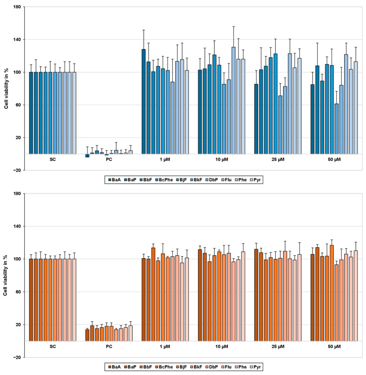
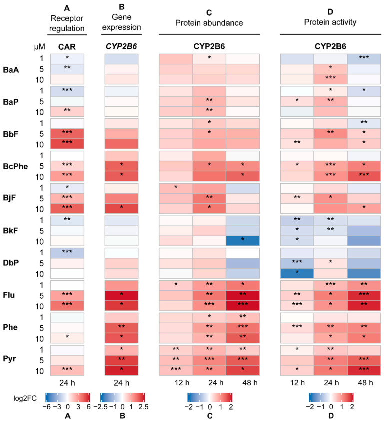
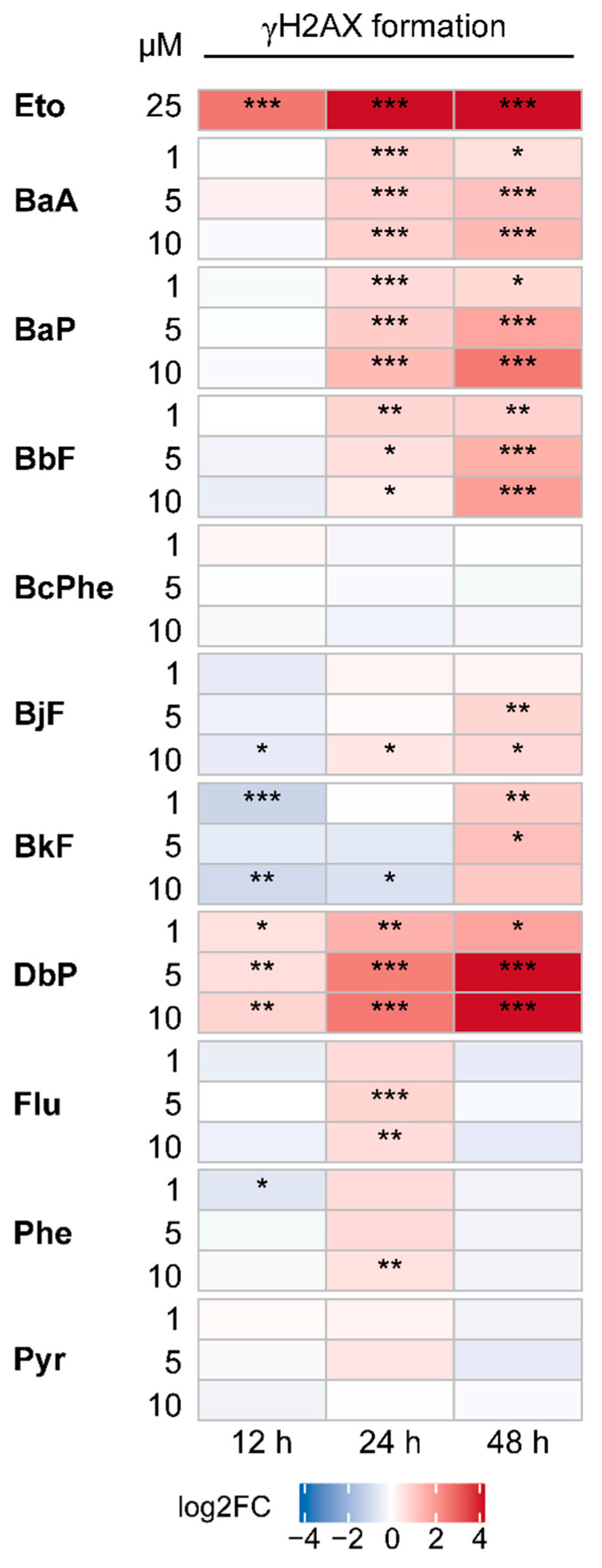
Polycyclic aromatic hydrocarbons (PAHs) are environmental pollutants produced by incomplete combustion of organic matter. They induce their own metabolism by upregulating xenobiotic-metabolizing enzymes such as cytochrome P450 monooxygenase 1A1 (CYP1A1) by activating the aryl hydrocarbon receptor (AHR). However, previous studies showed that individual PAHs may also interact with the constitutive androstane receptor (CAR). Here, we studied ten PAHs, different in carcinogenicity classification, for their potential to activate AHR- and CAR-dependent luciferase reporter genes in human liver cells. The majority of investigated PAHs activated AHR, while non-carcinogenic PAHs tended to activate CAR. We further characterized gene expression, protein abundancies and activities of the AHR targets CYP1A1 and 1A2, and the CAR target CYP2B6 in human HepaRG hepatoma cells. Enzyme induction patterns strongly resembled the profiles obtained at the receptor level, with AHR-activating PAHs inducing CYP1A1/1A2 and CAR-activating PAHs inducing CYP2B6. In summary, this study provides evidence that beside well-known activation of AHR, some PAHs also activate CAR, followed by subsequent expression of respective target genes. Furthermore, we found that an increased PAH ring number is associated with AHR activation as well as the induction of DNA double-strand breaks, whereas smaller PAHs activated CAR but showed no DNA-damaging potential.

Keywords: liver; nuclear receptors; polycyclic aromatic hydrocarbons; toxicity; xenobiotic metabolism.


Conflict of interest statement

The authors declare the following competing financial interest: Oliver Poetz is a shareholder of SIGNATOPE GmbH. SIGNATOPE offers assay development and service using MS-based immunoassay technology.

Figures

Figure 1
Figure 1
Chemical structures of polycyclic aromatic hydrocarbons (PAHs) used in this study in alphabetical order. PAHs are clustered additionally according to their International Agency for Research on Cancer (IARC) classification. Group (1): carcinogenic to humans; group (2A): probably carcinogenic to humans; group (2B): possibly carcinogenic to humans; group (3): not classifiable as to its carcinogenicity to humans [22].
Figure 2
Figure 2
Cell viability of HepG2 (blue) and HepaRG cells (orange) 24 h and 48 h after polycyclic aromatic hydrocarbon (PAH) treatment, respectively. To determine cell viability, the 3-(4,5-dimethylthiazol-2-yl)-2,5-diphenyltetrazolium bromide (MTT) assay was performed. After treatment with the test compounds, 10 µL MTT (5 mg/mL in PBS) was added to each well and incubated for 2 h (37 °C, 5% CO2). To dissolve the blue-colored formazan crystals, incubation medium was exchanged for 130 µL/well pre-warmed desorption solution (0.7% sodium dodecyl sulphate (SDS) in propan-2-ol) and gently shaken for 15 min. Afterward, absorbance was measured at 590 nm on a Tecan Infinite M200 Pro spectrophotometer (Tecan group, Männedorf, Switzerland). Obtained data were normalized to the solvent control (SC); 0.01% Triton-X 100 served as a positive control (PC). Results are expressed as mean + SD, n = 3.
Figure 3
Figure 3
Cumulated heatmaps of receptor regulation (A), gene expression (B), protein abundance (C), and protein activity (D) of aryl hydrocarbon receptor (AHR) and its targets cytochrome P450 monooxygenase 1A1 (CYP1A1) and 1A2 (CYP1A2), respectively. Respective experiments were conducted as described in the Materials and Methods section. For protein abundance and activity, the following applied: For values below the respective lower limit of quantification (LLOQ), the value half of the LLOQ was used for calculations. Generally, mean values were calculated from the individually measured raw data. Fold changes with regard to the respective solvent control were determined and then log2-transformed (log2FC). A positive regulation is indicated by red shades, a negative regulation by blue shades. Statistical analysis was performed with Student’s t-test (n = 3, * p ≤ 0.05, ** p ≤ 0.01, *** p ≤ 0.001). Underlying fold changes and standard deviations can be found in the Supplementary Materials “Underlying Data”.
Figure 4
Figure 4
Cumulated heatmaps of receptor regulation (A), gene expression (B), protein abundance (C), and protein activity (D) of constitutive androstane receptor (CAR) and its target cytochrome P450 monooxygenase 2B6 (CYP2B6). Respective experiments were conducted as described in the Materials and Methods section. For protein abundance and activity, the following applied: For values below the respective lower limit of quantification (LLOQ), the value half of the LLOQ was used for calculations. Generally, mean values were calculated from the individually measured raw data. Fold changes with regard to the respective solvent control were determined and then log2-transformed (log2FC). A positive regulation is indicated by red shades, a negative regulation by blue shades. Statistical analysis was performed with Student’s t-test (n = 3, * p ≤ 0.05, ** p ≤ 0.01, *** p ≤ 0.001). Underlying fold changes and standard deviations can be found in in the Supplementary Materials “Underlying Data”.
Figure 5
Figure 5
Frequency of histone H2AX phosphorylation. HepaRG cells were treated with 1, 5, or 10 µM of each PAH and incubated for 12, 24, 48 h. Etoposide (Eto; 25 µM) served as a metabolism-independent positive control. Afterward, cells were fixed, blocked, and stained with a combination of primary anti-γH2AX and secondary Alexa Fluor 647-antibody (red). Nuclei were counterstained using 4′,6-diamidino-2-phenylindole (DAPI) (blue). The figure depicts representative fluorescence images (brightness +30%) of each channel as well as a merged picture, respectively, 48 h posttreatment with solvent control (SC), Eto, or 10 µM PAH. Images were taken with Celldiscoverer (Zeiss, Oberkochen, Germany) and Zeiss Blue software (Zen 3.1 blue edition, Zeiss, Oberkochen, Germany).
Figure 6
Figure 6
Quantitative analysis of H2AX phosphorylation presented as heatmap. Based on fluorescence images, fluorescence intensity was quantified. Following, fold induction over DAPI signal was calculated and referred to solvent control. Data were then log2-transformed (log2FC). An increase in H2AX phosphorylation is indicated by red shades, a decrease by blue shades. Statistical analysis was performed with Student’s t-test (n = 3, * p ≤ 0.05, ** p ≤ 0.01, *** p ≤ 0.001). Fold changes and standard deviations can be found in in the Supplementary Materials “Underlying Data”.

References

    1. Boström C.E., Gerde P., Hanberg A., Jernstrom B., Johansson C., Kyrklund T., Rannug A., Tornqvist M., Victorin K., Westerholm R. Cancer risk assessment, indicators, and guidelines for polycyclic aromatic hydrocarbons in the ambient air. Environ. Health Perspect. 2002;110(Suppl. S3):451–488. doi: 10.1289/ehp.110-1241197. - DOI - PMC - PubMed
    1. IARC Some non-heterocyclic polycyclic aromatic hydrocarbons and some related exposures. Monogr. Eval. Carcinog. Risks Hum. 2010;92:1. - PMC - PubMed
    1. Shimada T. Xenobiotic-metabolizing enzymes involved in activation and detoxification of carcinogenic polycyclic aromatic hydrocarbons. Drug Metab. Pharmacokinet. 2006;21:257–276. doi: 10.2133/dmpk.21.257. - DOI - PubMed
    1. Koreeda M., Moore P.D., Wislocki P.G., Levin W., Yagi H., Jerina D.M. Binding of benzo[a]pyrene 7,8-diol-9,10-epoxides to DNA, RNA, and protein of mouse skin occurs with high stereoselectivity. Science. 1978;199:778–781. doi: 10.1126/science.622566. - DOI - PubMed
    1. Xue W., Warshawsky D. Metabolic activation of polycyclic and heterocyclic aromatic hydrocarbons and DNA damage: A review. Toxicol. Appl. Pharmcol. 2005;206:73–93. doi: 10.1016/j.taap.2004.11.006. - DOI - PubMed

MeSH terms

Substances