

Exosomal miRNAs as Potential Biomarkers to Monitor Phosphodiesterase 5 Inhibitor Induced Anti-Fibrotic Effects on CCl4 Treated Rats
- PMID: 33396535
- PMCID: PMC7795540
- DOI: 10.3390/ijms22010382
Exosomal miRNAs as Potential Biomarkers to Monitor Phosphodiesterase 5 Inhibitor Induced Anti-Fibrotic Effects on CCl4 Treated Rats
Abstract
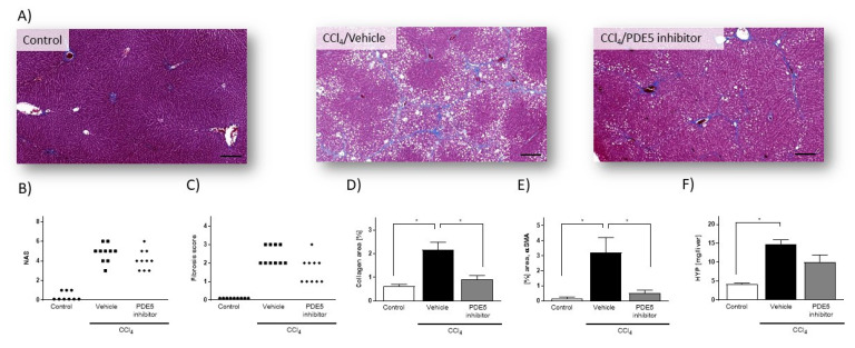
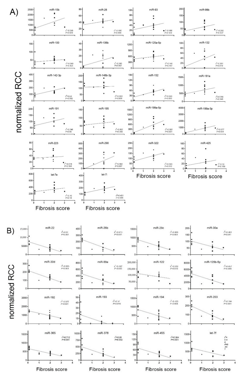
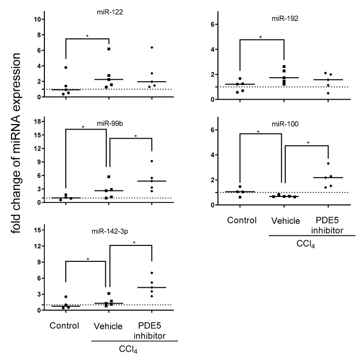
MicroRNAs (miRNAs) are short, non-coding RNA species that are important post-transcriptional regulators of gene expression and play an important role in the pathogenesis of non-alcoholic fatty liver disease. Here, we investigated the phosphodiesterase 5 (PDE5) inhibitor induced effects on hepatic and plasma exosomal miRNA expression in CCl4-treated rats. In the present study, hepatic miRNA profiling was conducted using the Nanostring nCounter technology and mRNA profiling using RNA sequencing from PDE5 treated rats in the model of CCl4-induced liver fibrosis. To evaluate if the PDE5 inhibitor affected differentially expressed miRNAs in the liver can be detected in plasma exosomes, qRT-PCR specific assays were used. In livers from CCl4-treated rats, the expression of 22 miRNAs was significantly increased (> 1.5-fold, adj. p < 0.05), whereas the expression of 16 miRNAs was significantly decreased (> 1.5-fold, adj. p < 0.05). The majority of the deregulated miRNA species are implicated in fibrotic and inflammatory processes. The PDE5 inhibitor suppressed the induction of pro-fibrotic miRNAs, such as miR-99b miR-100 and miR-199a-5p, and restored levels of anti-fibrotic miR-122 and miR-192 in the liver. In plasma exosomes, we observed elevated levels of miR-99b, miR-100 and miR-142-3p after treatment with the PDE5-inhibitor compared to CCl4/Vehicle-treated. Our study demonstrated for the first time that during the development of hepatic fibrosis in the preclinical model of CCl4-induced liver fibrosis, defined aspects of miRNA regulated liver pathogenesis are influenced by PDE5 treatment. In conclusion, miRNA profiling of plasma exosomes might be used as a biomarker for NASH progression and monitoring of treatment effects.
Keywords: CCl4; PDE 5 inhibitor; exosomes; gene expression; liver fibrosis; microRNAs.
Conflict of interest statement
The authors declare no conflict of interest.
Figures


References
-
- Chalasani N., Younossi Z., Lavine J.E., Diehl A.M., Brunt E.M., Cusi K., Charlton M., Sanyal A.J. American Gastroenterological Association; American Association for the Study of Liver Diseases; American College of Gastroenterology. The diagnosis and management of non-alcoholic fatty liver disease: Practice guideline by the American Gastroenterological Association, American Association for Study of Liver Diseases, and American College of Gastroenterology. Gastroenterology. 2012;142:1592–1609. - PubMed
MeSH terms
Substances
LinkOut - more resources
Full Text Sources
Medical
