. 2020 Dec 30;10(1):101.
doi: 10.3390/jcm10010101.

Vertical Ridge Augmentation of Fibula Flap in Mandibular Reconstruction: A Comparison between Vertical Distraction, Double-Barrel Flap and Iliac Crest Graft

Affiliations

Vertical Ridge Augmentation of Fibula Flap in Mandibular Reconstruction: A Comparison between Vertical Distraction, Double-Barrel Flap and Iliac Crest Graft

Carlos Navarro Cuéllar et al. J Clin Med. .

Abstract

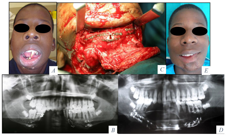
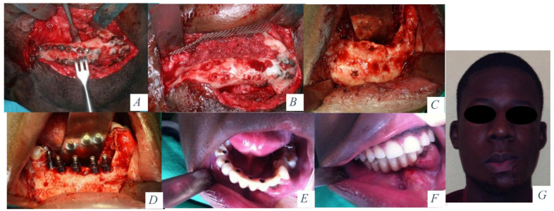
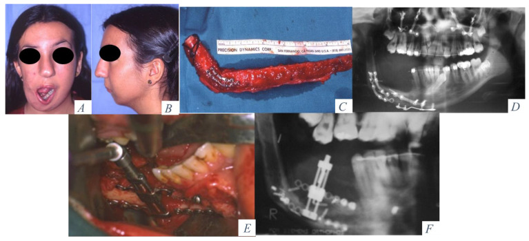
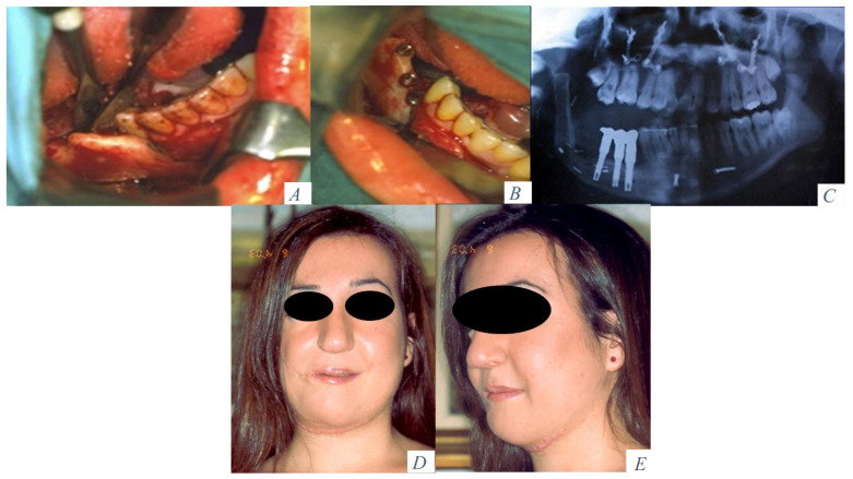
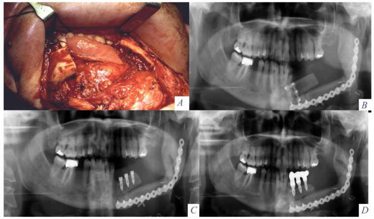
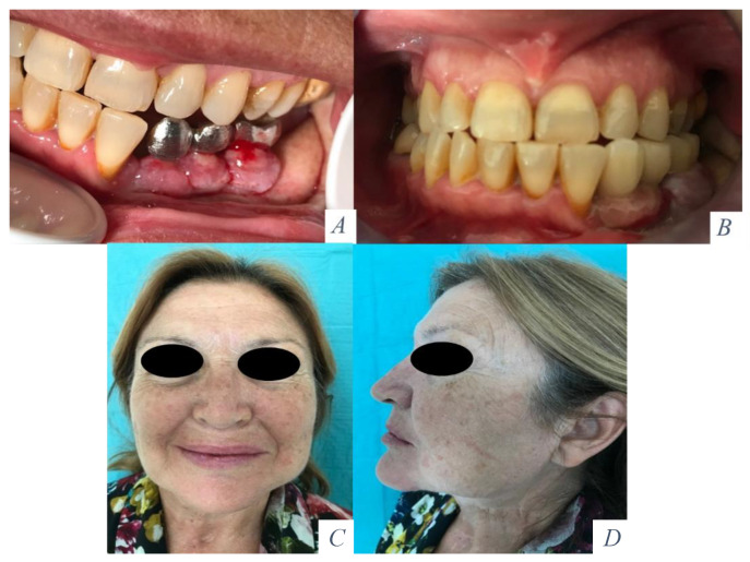
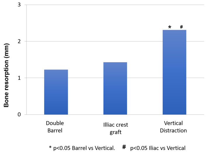
Double-barrel flap, vertical distraction and iliac crest graft are used to reconstruct the vertical height of the fibula. Twenty-four patients with fibula flap were reconstructed comparing these techniques (eight patients in each group) in terms of height of bone, bone resorption, implant success rate and the effects of radiotherapy. The increase in vertical bone with vertical distraction, double-barrel flap and iliac crest was 12.5 ± 0.78 mm, 18.5 ± 0.5 mm, and 17.75 ± 0.6 mm, (p < 0.001). The perimplant bone resorption was 2.31 ± 0.12 mm, 1.23 ± 0.09 mm and 1.43 ± 0.042 mm (p < 0.001), respectively. There were significant differences in vertical bone reconstruction and bone resorption between double-barrel flap and vertical distraction and between iliac crest and vertical distraction (p < 0.001). The study did not show significant differences in implant failure (p = 0.346). Radiotherapy did not affect vertical bone reconstruction (p = 0.125) or bone resorption (p = 0.237) but it showed higher implant failure in radiated patients (p = 0.015). The double-barrel flap and iliac crest graft showed better stability in the height of bone and less bone resorption and higher implant success rates compared with vertical distraction. Radiation therapy did not affect the vertical bone reconstruction but resulted in a higher implant failure.

Keywords: double-barrel flap; fibula flap; iliac crest graft; vertical augmentation; vertical distraction.


Conflict of interest statement

The authors declare no conflict of interest.

Figures

Figure 1
Figure 1
(A) Labial incompetence, salivary incontinence, impossibility to accomplish a normal diet and speech. (B) Segmental bone defect at the symphysis and right mandibular body. (C) Fibula osteocutaneous flap for composite reconstruction. Semirigid fixation. (D) Panoramic radiograph after mandibular reconstruction. (E) Recovery of mandibular symmetry, oral opening and oral competence.
Figure 2
Figure 2
(A) Cervical approach and removal of the osteosynthesis material. (B) Corticocancellous iliac crest bone graft and titanium mesh. (C) Mandibular height and width gained with the graft 6 months later. (D) Placement of 5 implants (Ticare) in the neomandible. (E) Prosthetic rehabilitation with a fixed implant supported prosthesis. (F) Final occlusion. (G) Final aesthetic result.
Figure 3
Figure 3
(A) Hemimandibular agenesis with facial asymmetry and mandibular deviation. (B) Retrusion of the lower facial third. (C) Mandibular reconstruction with an osseous fibula flap. (D) Panoramic radiograph with vertical discrepancy between the remaining mandible and the fibula flap. Le Fort I osteotomy for occlusal compensation. (E) Alveolar distractor placed in the fibula. (F) Panoramic radiograph at the beginning of the distraction procedure.
Figure 4
Figure 4
(A) Height and width of bone obtained after alveolar distraction. (B) Placement of 3 dental implants in the fibula flap. (C) Prosthetic rehabilitation with a fixed implant-supported prosthesis. (D) Facial symmetry and aesthetic result after mandibular reconstruction and oral rehabilitation. (E) Projection of the lower third face.
Figure 5
Figure 5
(A) Squamous cell carcinoma of the floor of the mouth with mandibular invasion. Tumor resection with segmental mandibulectomy and clear margins. (B) Immediate reconstruction with a double-barrel osseocutaneous free flap. Rigid fixation between the lower layer of the fibula and the remaining mandible. Semirigid fixation between the upper layer and the mandible. (C) Removal of part of the osteosynthesis material and placement of three implants (Ticare). (D) Prosthetic rehabilitation with a fixed implant supported prosthesis.
Figure 6
Figure 6
(A) Metal framework evaluated intraorally. (B) Functional rehabilitation with a ceramic fixed implant supported prosthesis. (C) Aesthetic result with mandibular symmetry. (D) Aesthetic profile with a good projection of the lower facial third.
Figure 7
Figure 7
Mann-Whitney analysis for vertical bone reconstruction.
Figure 8
Figure 8
Mann-Whitney analysis for bone resorption.

References

    1. Cuellar C.N., Gil M.C., Delgado J.P., Martínez B.G., Sanz J.A., de Atalaya J.L., Ochandiano S., Vila C.N. Reconstrucción oromandibular con colgajo libre de peroné e implantes osteointegrados. Acta Otorrinolaringol. Esp. 2003;54:54–64. doi: 10.1016/S0001-6519(03)78384-8. - DOI - PubMed
    1. He Y., Zhang Z.Y., Zhu H.G., Wu Y.Q., Fu H.H. Double-Barrel fibula vascularized free flap with dental rehabilitation for mandibular reconstruction. J. Oral Maxillofac. Surg. 2011;69:2663–2669. doi: 10.1016/j.joms.2011.02.051. - DOI - PubMed
    1. Shen Y., Sun J., Mei-mei L., Huang W., Ow A. Special considerations in virtual surgical planning for secondary accurate maxillary reconstruction with vascularized fibula osteomyocutaneous flap. J. Plast. Reconstr. Aesthet. Surg. 2012;65:893–902. doi: 10.1016/j.bjps.2011.12.035. - DOI - PubMed
    1. Melville J.C., Manis C.S., Shum J.W., Alsuwied D. Single-Unit 3D- Printed Titanium reconstruction: The evolution of surgicasl reconstruction for maxillary defects—A case report and review of current techniques. J. Oral Maxillofac. Surg. 2019;77:874.e1–874.e13. doi: 10.1016/j.joms.2018.11.030. - DOI - PubMed
    1. Swendseid B., Roden D.F., Vimawala S., Richa T., Sweeny L., Goldman R.A., Luginbuhl A., Heffelfinger R.N., Khanna S., Curry J.M. Virtual Surgical Planning in Subscapular System Free Flap Reconstruction of midface defects. Oral Oncol. 2020;101:104508. doi: 10.1016/j.oraloncology.2019.104508. - DOI - PubMed

LinkOut - more resources