

Structural Aspects and Prediction of Calmodulin-Binding Proteins
- PMID: 33396740
- PMCID: PMC7795363
- DOI: 10.3390/ijms22010308
Structural Aspects and Prediction of Calmodulin-Binding Proteins
Abstract
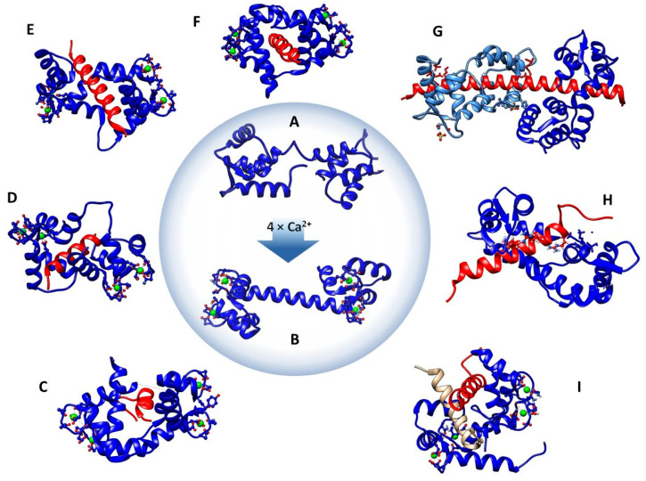
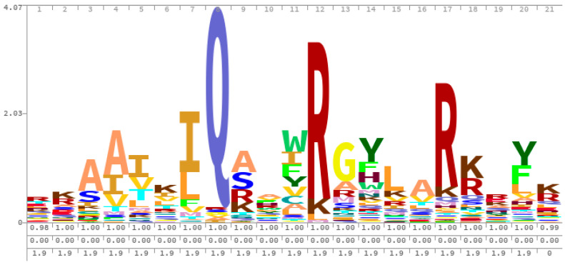
Calmodulin (CaM) is an important intracellular protein that binds Ca2+ and functions as a critical second messenger involved in numerous biological activities through extensive interactions with proteins and peptides. CaM's ability to adapt to binding targets with different structures is related to the flexible central helix separating the N- and C-terminal lobes, which allows for conformational changes between extended and collapsed forms of the protein. CaM-binding targets are most often identified using prediction algorithms that utilize sequence and structural data to predict regions of peptides and proteins that can interact with CaM. In this review, we provide an overview of different CaM-binding proteins, the motifs through which they interact with CaM, and shared properties that make them good binding partners for CaM. Additionally, we discuss the historical and current methods for predicting CaM binding, and the similarities and differences between these methods and their relative success at prediction. As new CaM-binding proteins are identified and classified, we will gain a broader understanding of the biological processes regulated through changes in Ca2+ concentration through interactions with CaM.
Keywords: CaMBP; IQ motif; SVM; calmodulin; machine learning; peptide; prediction; random forest.
Conflict of interest statement
The authors declare no conflict of interest.
Figures
References
-
- Nyegaard M., Overgaard M.T., Sondergaard M.T., Vranas M., Behr E.R., Hildebrandt L.L., Lund J., Hedley P.L., Camm A.J., Wettrell G., et al. Mutations in calmodulin cause ventricular tachycardia and sudden cardiac death. Am. J. Hum. Genet. 2012;91:703–712. doi: 10.1016/j.ajhg.2012.08.015. - DOI - PMC - PubMed
-
- Makita N., Yagihara N., Crotti L., Johnson C.N., Beckmann B.M., Roh M.S., Shigemizu D., Lichtner P., Ishikawa T., Aiba T., et al. Novel calmodulin mutations associated with congenital arrhythmia susceptibility. Circ. Cardiovasc. Genet. 2014;7:466–474. doi: 10.1161/CIRCGENETICS.113.000459. - DOI - PMC - PubMed
-
- Kawasaki H., Nakayama S., Kretsinger R.H. Classification and evolution of EF-hand proteins. Biometals Int. J. Role Met. Ions Biol. Biochem. Med. 1998;11:277–295. - PubMed
Publication types
MeSH terms
Substances
LinkOut - more resources
Full Text Sources
Research Materials
Miscellaneous
