Clinical Trial
. 2021 Apr;32(7-8):375-389.
doi: 10.1089/hum.2019.255. Epub 2021 Feb 18.

Dose-Escalation Study of Systemically Delivered rAAVrh74.MHCK7.micro-dystrophin in the mdx Mouse Model of Duchenne Muscular Dystrophy

Affiliations
Clinical Trial

Dose-Escalation Study of Systemically Delivered rAAVrh74.MHCK7.micro-dystrophin in the mdx Mouse Model of Duchenne Muscular Dystrophy

Rachael A Potter et al. Hum Gene Ther. 2021 Apr.

Abstract

Duchenne muscular dystrophy (DMD) is a rare, X-linked, fatal, degenerative neuromuscular disease caused by mutations in the DMD gene. More than 2,000 mutations of the DMD gene are responsible for progressive loss of muscle strength, loss of ambulation, and generally respiratory and cardiac failure by age 30. Recently, gene transfer therapy has received widespread interest as a disease-modifying treatment for all patients with DMD. We designed an adeno-associated virus vector (rAAVrh74) containing a codon-optimized human micro-dystrophin transgene driven by a skeletal and cardiac muscle-specific promoter, MHCK7. To test the efficacy of rAAVrh74.MHCK7.micro-dystrophin, we evaluated systemic injections in mdx (dystrophin-null) mice at low (2 × 10¹² vector genome [vg] total dose, 8 × 10¹³ vg/kg), intermediate (6 × 10¹² vg total dose, 2 × 10¹⁴ vg/kg), and high doses (1.2 × 10¹³ vg total dose, 6 × 10¹⁴ vg/kg). Three months posttreatment, specific force increased in the diaphragm (DIA) and tibialis anterior muscle, with intermediate and high doses eliciting force outputs at wild-type (WT) levels. Histological improvement included reductions in fibrosis and normalization of myofiber size, specifically in the DIA, where results for low and intermediate doses were not significantly different from the WT. Significant reduction in central nucleation was also observed, although complete normalization to WT was not seen. No vector-associated toxicity was reported either by clinical or organ-specific laboratory assessments or following formal histopathology. The findings in this preclinical study provided proof of principle for safety and efficacy of systemic delivery of rAAVrh74.MHCK7.micro-dystrophin at high vector titers, supporting initiation of a Phase I/II safety study in boys with DMD.

Keywords: AAV; Duchenne muscular dystrophy; dose-escalation; gene therapy; mdx mouse model; micro-dystrophin; rAAVrh74.MHCK7.micro-dystrophin.


Conflict of interest statement

R.A.P. is an employee of Sarepta Therapeutics, Inc., D.A.G. is an employee of Sarepta Therapeutics, Inc., K.N.H. and E.K.C.: No competing financial interest exists. E.L.P. is an employee of Sarepta Therapeutics, Inc., J.R.M. is the coinventor of the rAAVrh74.micro-dystrophin technology. This technology has been exclusively licensed to Sarepta Therapeutics, Inc., L.R.R.-K. is the coinventor of the AAVrh74.micro-dystrophin technology and eligible to receive financial consideration as a result. L.R.R.-K. is an employee of Sarepta Therapeutics, Inc.

Figures
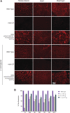
Figure 1.
Figure 1.
Robust micro-dystrophin expression at the sarcolemma membrane of skeletal and cardiac muscle after systemic delivery of rAAVrh74.MHCK7.micro-dystrophin. Mice received rAAVrh74.MHCK7.micro-dystrophin intravenously at 2.0 × 1012 vector genome (vg) total (low), 6 × 1012 vg total (intermediate), and 1.2 × 1013 vg total (high) dose. (A) Representative immunohistochemistry images are shown at the intermediate dose of 6 × 1012 vg total. Immunofluorescent staining for micro-dystrophin using an N-terminal dystrophin antibody in the TA, heart, and DIA 3 months postinjection compared to Lactated Ringer's (vehicle)-treated mdx mice (second row panel). β-sarcoglycan staining in the treated cohorts represent rescue of the dystrophin-associated protein complex. (B) Quantification of immunofluorescent-positive fibers expressing dystrophin protein. Data are reported as mean ± SEM of n = 5 per treatment group. DIA, diaphragm; GAS, gastrocnemius; GLUT, gluteus; mdx-LR, Lactated Ringer's (vehicle)-treated mdx; PSO, psoas major; QD, quadriceps; SEM, standard error of the mean; TA, tibialis anterior; TRI, triceps.
Figure 2.
Figure 2.
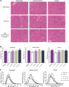
Systemic treatment with rAAVrh74.MHCK7.micro-dystrophin improves muscle pathology in mdx mice. Hematoxylin and eosin staining to detect morphology. (A) Representative images of diaphragm, tibialis anterior, and triceps muscle from WT mice, Lactated Ringer's (vehicle)-treated mdx mice (mdx-LR mice), and rAAVrh74.MHCK7.micro-dystrophin-treated mdx mice at the intermediate dose of 6 × 1012 vg total. (B, C) Quantification of average fiber size and frequency distribution demonstrates a normalization of fiber size across all of the assessed tissues (i.e., diaphragm, tibialis anterior, and triceps) in the mdx mice treated with rAAVrh74.MHCK7.micro-dystrophin at total dose of 2 × 1012 vg, 6 × 1012 vg, and 1.2 × 1013 vg. Data reported in bar graph as mean ± SEM (n = 4 low dose (SDDIA = 7.4, SDTA = 15.1, SDTRI = 17.3); n = 8 intermediate dose (SDDIA = 7.2, SDTA = 14.1, SDTRI = 15.2) and high dose (SDDIA = 7.9, SDTA = 12, SDTRI = 15.8); n = 6 mdx-LR (SDDIA = 6.6, SDTA = 15.8, SDTRI = 15.1); and WT (SDDIA = 5.6, SDTA = 10, SDTRI = 10.6)). Data were analyzed by one-way ANOVA followed by Tukey's post hoc analysis. **** = p < 0.0001 versus mdx-LR mice. + = p < 0.5; ++ = p < 0.005; ++++ = p < 0.0001 versus WT. ANOVA, analysis of variance; SD, standard deviation; WT, wild-type.
Figure 3.
Figure 3.
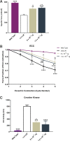
Reduction of fibrosis after systemic treatment with rAAVrh74.MHCK7.micro-dystrophin is associated with improvement in DIA muscle function. Muscle tissue sections were stained using picrosirius red stain to assess collagen content. (A) Representative images of DIA tissue sections in WT mice, Lactated Ringer's (vehicle)-treated mdx mice (mdx-LR mice), and mdx mice treated with rAAVrh74.MHCK7.micro-dystrophin with low (2 × 1012 vg), intermediate (6 × 1012 vg), and high (1.2 × 1013 vg) doses. (B) Quantification of collagen accumulation (%) in the DIA of WT mice, mdx-LR mice, and mdx mice treated with rAAVrh74.MHCK7.micro-dystrophin (n = 5, low dose; n = 8, intermediate and high dose; n = 6, mdx-LR; and n = 5, WT). (C) DIA muscle function in WT mice, mdx-LR mice, and mdx mice treated with rAAVrh74.MHCK7.micro-dystrophin (n = 5, low dose; n = 9, intermediate; n = 8, high dose; n = 8, mdx-LR; and n = 5, WT). Following 3 months of treatment, DIA muscle strips were harvested to measure specific force (normalized to cross-sectional area). For panels B and C, data are represented as mean ± SEM and were analyzed by one-way ANOVA followed by Tukey's post hoc analysis. ** = p < 0.005 versus mdx-LR mice; + = p < 0.05, ++ = p < 0.005, +++ = p < 0.001 versus WT mice.
Figure 4.
Figure 4.
Functional benefits to skeletal muscle in intermediate- and high-dose cohorts. (A) Specific muscle force in TA muscles (normalized to cross-sectional area) assessed 3 months after treatment with rAAVrh74.MHCK7.micro-dystrophin (left and right side per animal, per cohort: n = 8, intermediate dose (6 × 1012 vg); n = 16, high dose (1.2 × 1013 vg); n = 14, Lactated Ringer's (vehicle)-treated mdx (mdx-LR); n = 9, WT). (B) Eccentric force loss in TA muscle assessed using a rigorous ECC protocol. Force output is plotted at each cycle (mean of left and right side per animal, per cohort: n = 8, intermediate dose; n = 13, high dose; n = 13, mdx-LR; n = 9, WT). (C) Serum CK levels 3 months post-treatment (n = 2, intermediate and high dose; n = 5, mdx-LR and WT). For panels A and C, data are represented as mean ± SEM and analyzed by one-way ANOVA followed by Tukey's post hoc analysis for multiple comparisons. ** = p < 0.01, *** = p < 0.001, and **** = p < 0.0001 versus mdx-LR mice; + = p < 0.05, ++ = p < 0.005, +++ = p < 0.001, ++++ = p = 0.0001 versus WT mice. In panel B, data are represented as mean ± SEM and analyzed by two-way ANOVA followed by Tukey's post hoc analysis for multiple comparisons. * = p < 0.05, ** = p < 0.01 versus mdx-LR mice; + = p < 0.05, +++ = p < 0.001, ++++ = p = 0.0001 versus WT mice. CK, creatine kinase; ECC, eccentric contraction cycle.
Figure 5.
Figure 5.
Biodistribution of vgs and protein expression in vector-treated mice. (A) Representative western blot of nNOS protein expression in muscle tissues of Lactated Ringer's (vehicle)-treated mdx mice, WT mice, and two mice (Mouse 347 and Mouse 346) treated with 6 × 1012 vg total dose of rAAVrh74.MHCK7.micro-dystrophin (intermediate). (B) Biodistribution of vgs normalized to microgram of DNA in organs and tissues after systemic delivery of rAAVrh74.MHCK7.micro-dystrophin (n = 3 for each cohort). Data are represented as mean ± SEM. (C) Western blot analysis of dystrophin protein expression in muscles and organs from mice 3 months after treatment with rAAVrh74.MHCK7.micro-dystrophin at an intermediate (6 × 1012 vg) and high (1.2 × 1013 vg) dose. α-actinin and γ-tubulin were used as loading controls. α-actin, α-actinin; Dys, dystrophin; γ-tub, γ-tubulin; Glut, gluteus; Gon, gonads; Hrt, heart; Kid, kidney; Liv, liver; Lng, lung; μDys, micro-dystrophin; mdx-LR, Lactated Ringer's (vehicle)-treated mdx; nNOS, neuronal nitric oxide synthase; Spl, spleen.
Figure 6.
Figure 6.
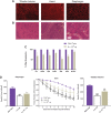
Long-term durability after a single administration of rAAVrh74.MHCK7.micro-dystrophin in mdx mice. (A, B) Representative immunofluorescent staining for micro-dystrophin using an N-terminal dystrophin antibody and hematoxylin and eosin stained TA, heart, and DIA of mdx mice treated with rAAVrh74.micro-dystrophin at an intermediate (6 × 1012 vg) dose, 6 months post-treatment. (C) Quantification of immunofluorescent-positive fibers expressing micro-dystrophin protein 6 months after a single treatment with rAAVrh74.MHCK7.micro-dystrophin. Data are reported as mean ± SEM of n = 5. (D) Specific force generation in the DIA and TA muscles 6 months after a single treatment with rAAVrh74.MHCK7.micro-dystrophin. Data reported as mean ± SEM of n = 5. Data were analyzed by one-way ANOVA followed by Tukey's post hoc analysis for multiple comparisons. ** = p < 0.05 versus mdx-LR mice; + = p < 0.05, ++ = p < 0.005, +++ = p < 0.0005, ++++ = p < 0.0001 versus WT mice. Eccentric force loss following a rigorous ECC protocol (n = 8 in each group). Data reported as mean ± SEM. Data were analyzed by two-way ANOVA followed by Tukey's post hoc analysis for multiple comparisons. + = p < 0.2, ++ = p < 0.007 versus WT mice. Glut, gluteus; mdx-LR, Lactated Ringer's (vehicle)-treated mdx.

References

    1. Aartsma-Rus A, Straub V, Hemmings R, et al. . Development of exon skipping therapies for Duchenne muscular dystrophy: a critical review and a perspective on the outstanding issues. Nucleic Acid Ther 2017;27:251–259 - PMC - PubMed
    1. Emery AE. Population frequencies of inherited neuromuscular disease—a world survey. Neuromuscul Disord 1991;1:19–29 - PubMed
    1. Mendell JR, Lloyd-Puryear M. Report of MDA muscle disease symposium on newborn screening for Duchenne muscular dystrophy. Muscle Nerve 2013;48:21–26 - PubMed
    1. Mendell JR, Shilling C, Leslie ND, et al. . Evidence based path to newborn screening for Duchenne muscular dystrophy. Ann Neurol 2012;71:304–313 - PubMed
    1. Oudet C, Hanauer A, Clemens P, et al. . Two hot spots of recombination in the DMD gene correlate with the deletion prone regions. Hum Mol Genet 1992;1:599–603 - PubMed

Publication types