. 2021 Jan 4;18(1):2.
doi: 10.1186/s12984-020-00764-5.

Dynamic changes of region-specific cortical features and scalp-to-cortex distance: implications for transcranial current stimulation modeling

Affiliations

Dynamic changes of region-specific cortical features and scalp-to-cortex distance: implications for transcranial current stimulation modeling

Hanna Lu et al. J Neuroeng Rehabil. .

Abstract

Background: Transcranial current stimulation in rehabilitation is a fast-growing field featured with computational and biophysical modeling. Cortical features and scalp-to-cortex distance (SCD) are key variables for determining the strength and distribution of the electric field, yet longitudinal studies able to capture these dynamic changes are missing. We sought to investigate and quantify the ageing effect on the morphometry and SCD of left primary motor cortex (M1) and dorsolateral prefrontal cortex (DLPFC) in normal ageing adults and mild cognitive impairment (MCI) converters.

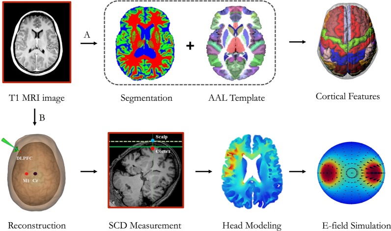
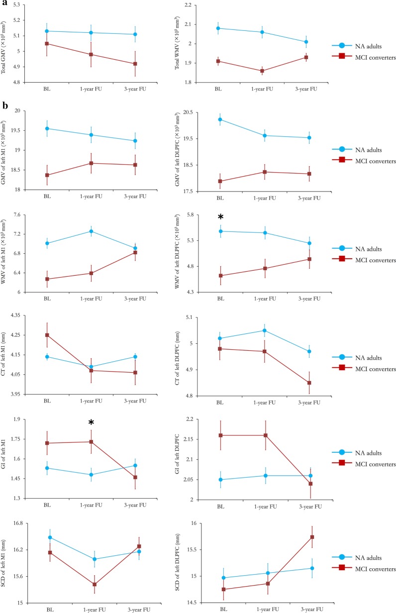
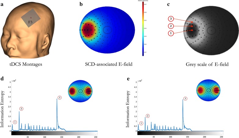
Methods: Baseline, 1-year and 3-year follow-up structural magnetic resonance imaging scans from normal ageing adults (n = 32), and MCI converters (n = 22) were drawn from the Open Access Series of Imaging Studies. We quantified the changes of the cortical features and SCDs of left M1 and DLPFC, including grey matter volume, white matter volume, cortical thickness, and folding. Head model was developed to simulate the impact of SCD on the electric field induced by transcranial current stimulation.

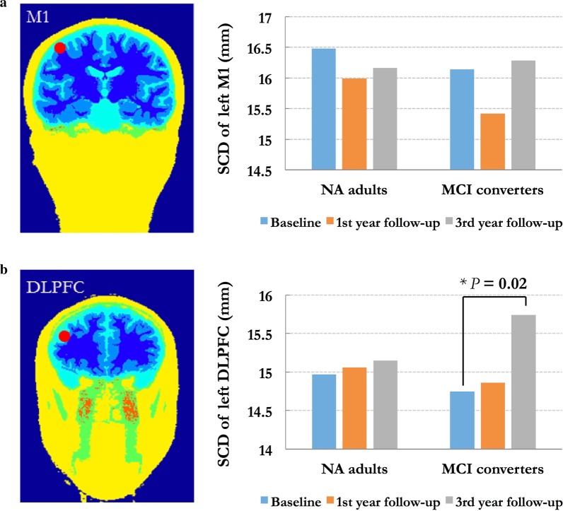
Results: Pronounced ageing effect was found on the SCD of left DLPFC in MCI converters. The SCD change of left DLPFC from baseline to 3-year follow-up demonstrated better performance to discriminate MCI converters from normal ageing adults than the other morphometric measures. The strength of electric field was consequently decreased with SCD in MCI converters.

Conclusion: Ageing has a prominent, but differential effect on the region-specific SCD and cortical features in older adults with cognitive impairments. Our findings suggest that SCD, cortical thickness, and folding of the targeted regions could be used as valuable imaging markers when conducting transcranial brain stimulation in individuals with brain atrophy.

Keywords: Ageing; Brain stimulation; Cortical folding; DLPFC; Modeling; Scalp-to-cortex distance.


Conflict of interest statement

The authors declare that they have no competing interests.

Figures

Fig. 1
Fig. 1
The pipeline of structural magnetic resonance imaging (sMRI) analysis: Path A represents the region-of-interest-based cortical mapping; Path B represents the measurement of scalp-to-cortex (SCD) and the construction of geometric simulation model
Fig. 2
Fig. 2
Trajectories demonstrating the region-specific morphometric measures listed in old adults with different cognitive statuses. In each plot, blue line represents the results from normal ageing (NA) adults, and red line represents the results from MCI converters. Error bars represent the standard error (SEM). * indicates significant between-group differences (p < 0.05)
Fig. 3
Fig. 3
Demonstration of region-specific scalp-to-cortex distance (SCD) in normal ageing (NA) adults and mild cognitive impairment (MCI) converters. a Importing individual structural MRI data to the neuronavigation system and constructing the scalp and cerebral cortex; b localizing the targets and measuring the SCD; c comparing the ageing effect on the SCD of left primary motor cortex (M1) in NA adults and MCI converters; d comparing the ageing effect on the SCD of left dorsolateral prefrontal cortex (DLPFC) in NA adults and MCI converters
Fig. 4
Fig. 4
Three-dimensional geometric simulation model of anodal transcranial current stimulation. a We first reconstructed the scalp and placed anodal electrode over left DLPFC (i.e., F3 in the international 10/20 system); b second, completed the simulation of SCD-associated E-field using SPHERES and ROAST toolbox, and c transferred the E-field to grey-scale images for entropy analysis. Finally, we calculated the entropy changes of simulated E-field in MCI converters at baseline (d) and 3-year follow-up (e)

Similar articles

Cited by

References

    1. Cole JH, Franke K. Predicting age using neuroimaging: innovative brain ageing biomarkers. Trends Neurosci. 2017;40:681–690. doi: 10.1016/j.tins.2017.10.001. - DOI - PubMed
    1. Chandra A, Dervenoulas G, Politis M. Alzheimer’s Disease Neuroimaging Initiative, Magnetic resonance imaging in Alzheimer’s disease and mild cognitive impairment. J Neurol. 2019;266:1293–1302. doi: 10.1007/s00415-018-9016-3. - DOI - PMC - PubMed
    1. Fjell AM, Westlye LT, Grydeland H, Amlien I, Espeseth T, Reinvang I, Initiative ADN. Accelerating cortical thinning: unique to dementia or universal in aging? Cereb Cortex. 2014;24:919–934. doi: 10.1093/cercor/bhs379. - DOI - PMC - PubMed
    1. Madan CR, Kensinger EA. Cortical complexity as a measure of age-related brain atrophy. NeuroImage. 2016;134:617–629. doi: 10.1016/j.neuroimage.2016.04.029. - DOI - PMC - PubMed
    1. Fjell AM, Walhovd KB, Fennema-Notestine C, McEvoy LK, Hagler DJ, Holland D, Dale AM. One-year brain atrophy evident in healthy aging. J Neurol. 2009;29:15223–15231. - PMC - PubMed

Publication types