. 2021 Jan 4;22(1):4.
doi: 10.1186/s13059-020-02222-w.

Integrated genomic and transcriptomic analysis reveals unique characteristics of hepatic metastases and pro-metastatic role of complement C1q in pancreatic ductal adenocarcinoma

Affiliations

Integrated genomic and transcriptomic analysis reveals unique characteristics of hepatic metastases and pro-metastatic role of complement C1q in pancreatic ductal adenocarcinoma

Jianyu Yang et al. Genome Biol. .

Abstract

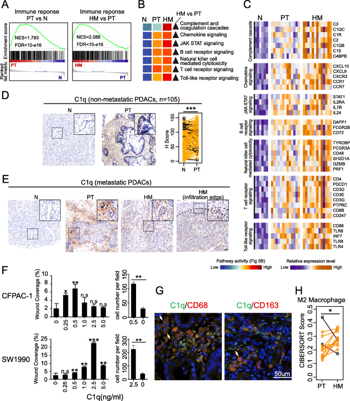
Background: Pancreatic ductal adenocarcinoma (PDAC) is one of the most lethal cancers due to its high metastasis rate in the liver. However, little is known about the molecular features of hepatic metastases due to difficulty in obtaining fresh tissues and low tumor cellularity.

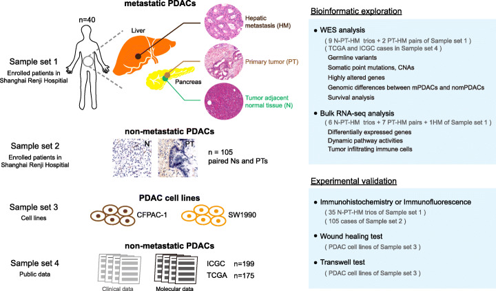
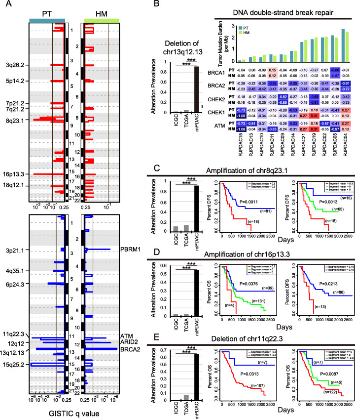
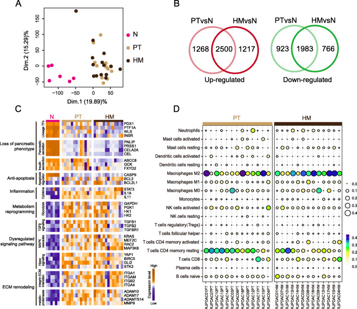
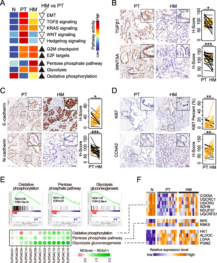
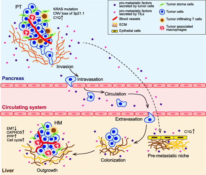
Results: We conduct exome sequencing and RNA sequencing for synchronous surgically resected primary tumors and the paired hepatic metastases from 17 hepatic oligometastatic pancreatic ductal adenocarcinoma and validate our findings in specimens from 35 of such cases. The comprehensive analysis of somatic mutations, copy number alterations, and gene expressions show high similarity between primary tumors and hepatic metastases. However, hepatic metastases also show unique characteristics, such as a higher degree of 3p21.1 loss, stronger abilities of proliferation, downregulation of epithelial to mesenchymal transition activity, and metabolic rewiring. More interesting, altered tumor microenvironments are observed in hepatic metastases, especially a higher proportion of tumor infiltrating M2 macrophage and upregulation of complement cascade. Further experiments demonstrate that expression of C1q increases in primary tumors and hepatic metastases, C1q is mainly produced by M2 macrophage, and C1q promotes migration and invasion of PDAC cells.

Conclusion: Taken together, we find potential factors that contribute to different stages of PDAC metastasis. Our study broadens the understanding of molecular mechanisms driving PDAC metastasis.

Keywords: C1q; Genomics; Hepatic metastasis; Pancreatic ductal adenocarcinoma; Transcriptomics; Tumor microenvironment.


Conflict of interest statement

The authors declare that they have no competing interests.

Figures

Fig. 1
Fig. 1
Study design and sample sets used in this study. Forty patients (sample set 1) were enrolled in this study. Genomic and transcriptomic profiling were performed to specimens from 17 patients of sample set 1. Experimental validation was performed using specimens from 35 patients of sample set 1, 105 non-metastatic patients from sample set 2, and two human PDAC cell lines from sample set 3. To explore the biological and clinical significance of identified events, molecular and survival data from previously published non-metastatic PDACs were used (sample set 4)
Fig. 2
Fig. 2
Potential clinical application of highly altered CNA events in hepatic metastatic PDAC. a Significantly occurred copy number amplifications (red, top panel) and deletions (blue, bottom panel) were identified in PT (left panel) and HM (right panel), respectively. Among them, deletion of 13q12.13 (b), amplification of 8q23.1 (c), amplification of 16q13.3 (d), and deletion of 11q22.3 (e) showed significantly higher alteration prevalence in metastatic PDACs (black bars in barplots) compared to non-metastatic PDACs (gray bars). ***P < 0.001. b Genes in chr13q12.13 contain BRCA2 that is a key component of DNA double-strand break repair pathway. Major components of this pathway showed recurrent CN loss in metastatic PDAC (right panel), suggesting these patients might be benefit from PPAR inhibitor. c Patients carrying CN gain of 8q23.1 are of higher metastatic risk. d, e CN amplification of 16q13.3 may serve as a biomarker for bad prognosis and higher metastatic risk while deletion of 11q22.3 may be used as a biomarker for good prognosis. As there was not enough case to test the association between DFS and gain of 16q13.3, we stratified patients according to a looser threshold. Patients with “segment_mean” of 16p13.3 greater than 0.15 were regarded as those with slight CN gain. DFS was defined as the interval between surgical treatment and date of diagnosis of distant metastases
Fig. 3
Fig. 3
HMs bear basic transcriptomic hallmarks of PDAC. a Principal component analysis based on whole transcriptome showed that HMs have similar transcriptome as PTs. As showed in first two principal components that HMs and PTs are closed clustered together. b Compared to Ns, there are many common DEGs shared by PTs and HMs, including 2500 upregulated and 1983 downregulated genes. c Similar as PTs, HMs showed basic molecular hallmarks including loss of pancreatic phenotype, anti-apoptosis, inflammation, metabolic remodeling, dysregulated signaling pathways, and ECM remodeling. Genes showed in heatmap were those common DEGs identified in b. d PTs and HMs shared similar tumor-infiltrating-lymphocytes profiles
Fig. 4
Fig. 4
Reversed EMT and metabolism rewiring in HMs. a Compared to PT, HMs showed decreased activities of EMT and EMT related pathways (upper panel) as well as increased cell cycle activity (bottom panel). This phenomenon was experimentally validated by IFH assays of 35 paired PTs and HMs. b HMs showed significant upregulation of epithelial marker E-cadherin and downregulation of mesenchymal marker N-cadherin. c TGFβ1 and WNT5A that are ligands of TGFβ signaling and WNT signaling, respectively, were significantly upregulated in HMs. d Proliferative marker Ki67 and CCNA2 were upregulated in HMs compare to PTs. *0.01 < P < 0.05; **0.001 < P < 0.01; ***P < 0.001. e Oxidative phosphorylation, pentose phosphate pathway, and glycolysis were up-regulated in HMs. Top panel shows results of between-group comparisons. Bottom panel shows difference between HMs and PTs of individual cases. Green indicates up regulation while purple indicates down regulation in HM. f Gene expression heat-map of key components of pathways showed in e
Fig. 5
Fig. 5
Tumor stroma plays important roles in PDAC metastasis. a Immune response was found gradually activated through N, PT, and HM. Comparing to Ns, the activity of immune response was increased in PTs (left panel). And it was further increased in HMs (right panel). b Multiple immune related pathways were found showing similar changing pattern as immune response. c Expression heatmap of representative genes of pathways showed in b. Among them, C1q that is involved in complement cascade was found mainly expressed at tumor stroma rather in tumor cells (d and e). Non-metastatic PDACs also showed overexpression of C1q in PTs compared to Ns (d). f Trans-well tests and wound healing test using PDAC cells lines CFPAC-1 and SW1990 showed that C1q would promote invasion and metastasis. g Identification of C1q-positive cells in PDAC. PDAC sections were double-stained for IF: C1q (green), CD68 (macrophage marker, red; left panel), CD163 (M2 macrophage marker, red; right panel). The double-positive cells appear in yellow (arrows). h In tumor microenvironment, M2 macrophages exhibited higher proportions in HMs compared to corresponding PTs
Fig. 6
Fig. 6
Schematic plot of multi-step hepatic metastasis of PDAC. To successfully build a second tumor clone, primary tumor cells of PDAC must go through a series of steps including local invasion, intravasation, surviving in the circulation system, extravasation, adapting to survival in new microenvironment, colonization, and outgrowth in liver. This figure was adapted from [43]. The types of cell that presented in tumor microenvironments and pro-metastatic factors marked in the figure were modified according to our findings

Similar articles

Cited by

References

    1. Rawla P, Sunkara T, Gaduputi V. Epidemiology of pancreatic cancer: global trends, etiology and risk factors. World J Oncol. 2019;10:10–27. doi: 10.14740/wjon1166. - DOI - PMC - PubMed
    1. Chandana S, Babiker HM, Mahadevan D. Therapeutic trends in pancreatic ductal adenocarcinoma (PDAC) Expert Opin Investig Drugs. 2019;28:161–177. doi: 10.1080/13543784.2019.1557145. - DOI - PubMed
    1. Siegel RL, Miller KD, Jemal A. Cancer statistics, 2019. CA Cancer J Clin. 2019;69:7–34. doi: 10.3322/caac.21551. - DOI - PubMed
    1. Yachida S, Iacobuzio-Donahue CA. The pathology and genetics of metastatic pancreatic cancer. Arch Pathol Lab Med. 2009;133:413–422. - PubMed
    1. Vincent A, Herman J, Schulick R, Hruban RH, Goggins M. Pancreatic cancer. Lancet. 2011;378:607–620. doi: 10.1016/S0140-6736(10)62307-0. - DOI - PMC - PubMed

Publication types

MeSH terms