Observational Study
. 2021 Jan 4;25(1):4.
doi: 10.1186/s13054-020-03440-1.

Protective reactive thymus hyperplasia in COVID-19 acute respiratory distress syndrome

Affiliations
Observational Study

Protective reactive thymus hyperplasia in COVID-19 acute respiratory distress syndrome

Pelagia Cuvelier et al. Crit Care. .

Abstract

Background: Patients with COVID-19 (COVID) may develop acute respiratory distress syndrome with or without sepsis, coagulopathy and visceral damage. While chest CT scans are routinely performed in the initial assessment of patients with severe pulmonary forms, thymus involvement and reactivation have not been investigated so far.

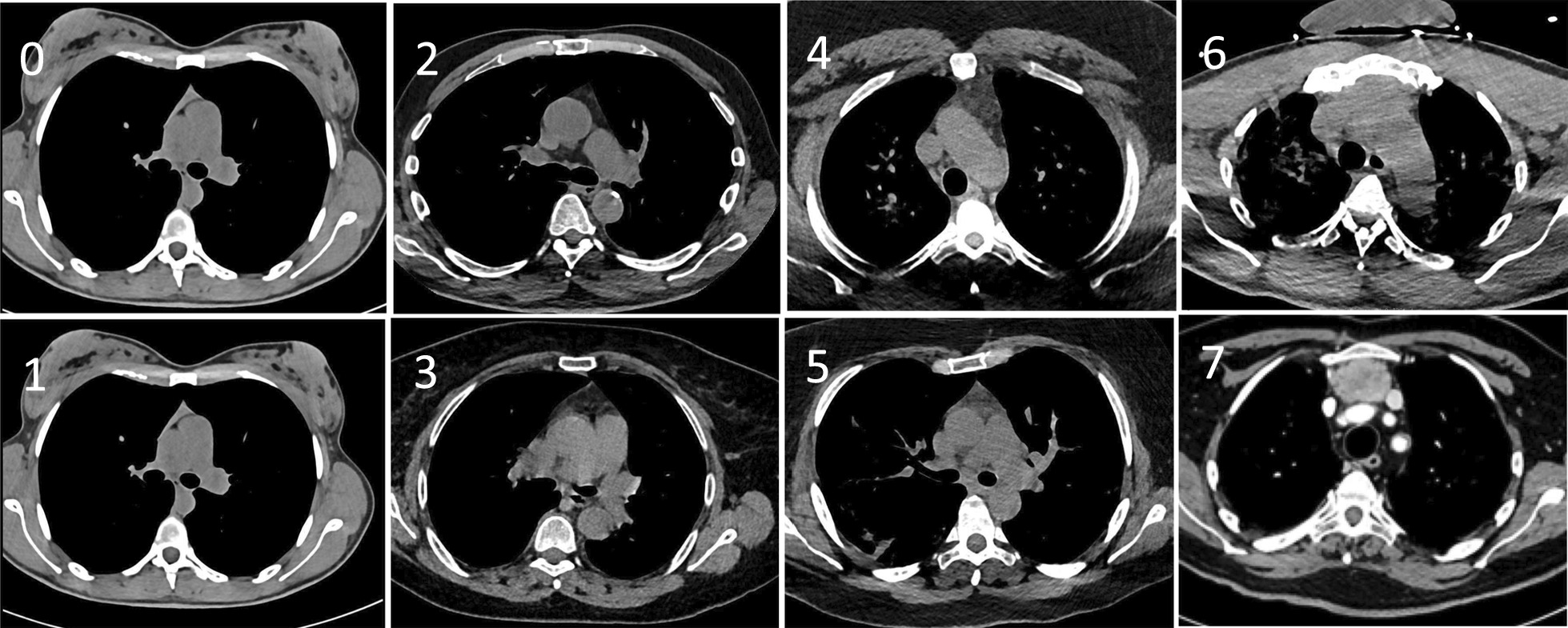
Methods: In this observational study, we systematically scored the enlargement of the thymus and the lung involvement, using CT scans, in all adult patients admitted to the ICU for COVID or any other cause (control group) at one centre between March and April 2020. Initial biological investigations included nasal detection of SARS-CoV-2 ribonucleic acid by polymerase chain reaction (PCR). In a subgroup of 24 patients with different degrees of pulmonary involvement and thymus hypertrophy, plasma cytokine concentrations were measured and the export of mature T cells from the thymus was estimated simultaneously by PCR quantification of T cell receptor excision circles (TRECs).

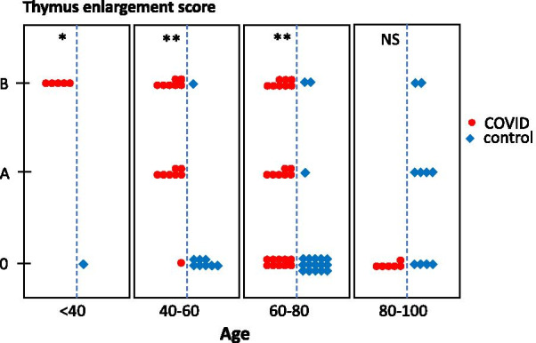
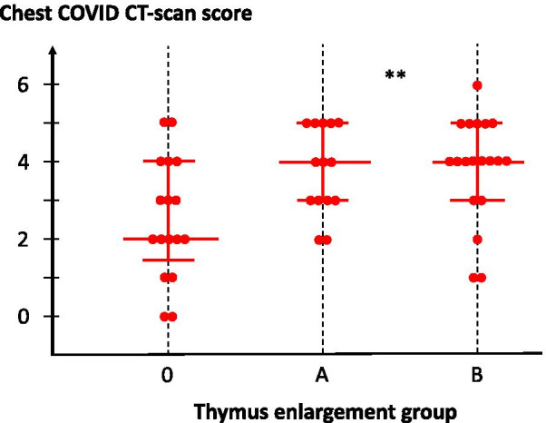
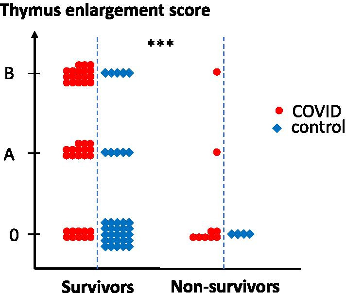
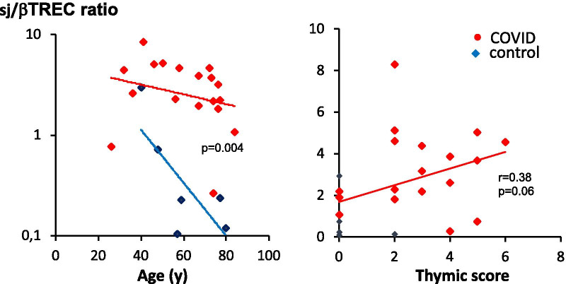
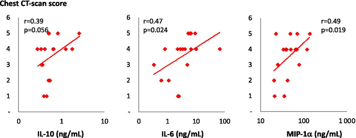
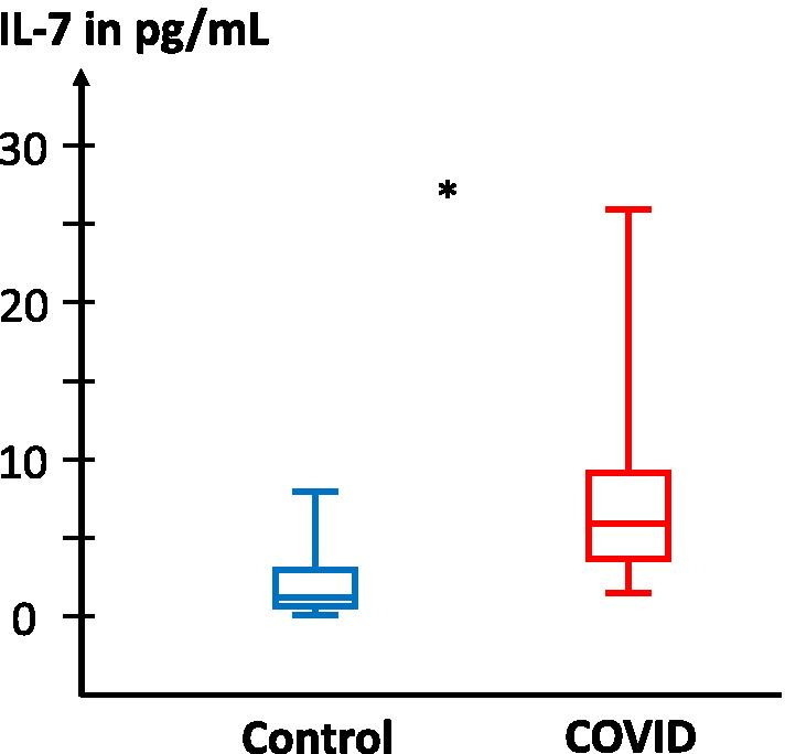
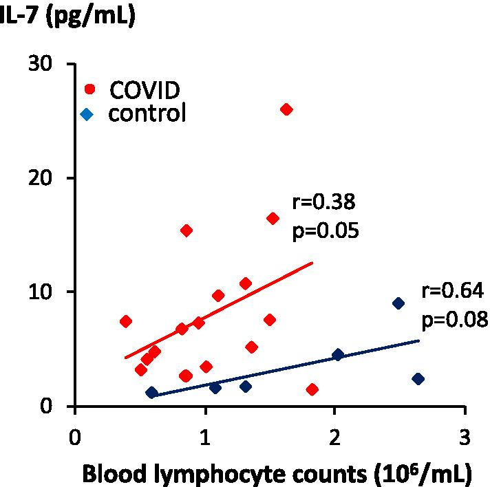
Results: Eighty-seven patients were studied: 50 COVID patients and 37 controls. Non-atrophic or enlarged thymus was more commonly observed in COVID patients than in controls (66% vs. 24%, p < 0.0001). Thymus enlargement in COVID patients was associated with more extensive lung injury score on CT scans (4 [3-5] vs. 2 [1.5-4], p = 0.01), but a lower mortality rate (8.6% vs. 41.2%, p < 0.001). Other factors associated with mortality were age, lymphopaenia, high CRP and co-morbidities. COVID patients had higher concentrations of IL-7 (6.00 [3.72-9.25] vs. 2.17 [1.76-4.4] pg/mL; p = 0.04) and higher thymic production of new lymphocytes (sj/βTREC ratio = 2.88 [1.98-4.51] vs. 0.23 [0.15-0.60]; p = 0.004). Thymic production was also correlated with the CT scan thymic score (r = 0.38, p = 0.03) and inversely correlated with the number of lymphocytes (r = 0.56, p = 0.007).

Conclusion: In COVID patients, thymus enlargement was frequent and associated with increased T lymphocyte production, which appears to be a beneficial adaptation to virus-induced lymphopaenia. The lack of thymic activity/reactivation in older SARS-CoV-2 infected patients could contribute to a worse prognosis.

Keywords: COVID-19; TREC; Thymus reactivation.


Conflict of interest statement

All authors declare no competing interests related to this study.

Figures

Fig. 1
Fig. 1
Chest CT scan images. Representative chest CT scan images of the thymic area of patients hospitalised for COVID, illustrating the classification of thymus aspects depicted in Table 2. Arrows indicate the different types of thymus hyperplasia
Fig. 2
Fig. 2
Thymic enlargement in COVID and control groups according to age categories. Thymic scores were determined through CT scan imaging in COVID (n = 50, red symbols) and COVID (n = 37, blue symbols) patients and displayed as a function of age categories. *p < 0.05; **p < 0.001, NS, not significant
Fig. 3
Fig. 3
Lung damage in COVID patients, according to thymus enlargement group. Lung CT scan score is presented for each patient, according to individual thymus CT scan. Each dot represents an individual patient. Median values with the 25–75% interquartile ranges are shown; **p = 0.01 between groups
Fig. 4
Fig. 4
Survival as a function of thymic CT scan score. Thymus CT scan score is presented for each patient in survivor and non-survivor groups. Statistical differences between groups are shown on top; ***p < 0.0001
Fig. 5
Fig. 5
Enhanced thymic function in COVID patients. The sj/βTREC ratio, which reflects the extent of thymic output, is represented as a function of age (left) or as a function of CT scan thymic score (right). Statistical differences between groups (ANCOVA using age as a concomitant variable), nonparametric correlation Pearson’s r and associated p value (one tailed) are indicated
Fig. 6
Fig. 6
Plasma concentrations of three cytokines as a function of chest CT scan score. Plasma concentrations of IL-10, IL-6 and MIP-1α were measured in patients hospitalised for COVID (n = 18) and are presented as a function of chest CT scan scores. Nonparametric correlation Pearson’s r and associated p value (one tail) are indicated
Fig. 7
Fig. 7
IL-7 concentrations in the plasma of control and COVID patients. IL-7 plasma concentration was measured in patients hospitalised for COVID (n = 18, red symbols) and controls (n = 6, blue symbols). Statistical significance of observed differences between groups are shown on top (Mann–Whitney U test)
Fig. 8
Fig. 8
IL-7 concentrations correlated with blood lymphocyte counts. IL-7 plasma concentration was measured in patients hospitalised for COVID (n = 18, red symbols) and controls (n = 6, blue symbols) and presented as a function of blood lymphocyte counts. Nonparametric correlation Pearson’s r and associated p value (one tail) are indicated

References

    1. Gandhi M, Yokoe DS, Havlir DV. Asymptomatic transmission, the Achilles’ heel of current strategies to control Covid-19. N Engl J Med. 2020;382:2158–2160. doi: 10.1056/NEJMe2009758. - DOI - PMC - PubMed
    1. Zhou F, Yu T, Du R, Fan G, Liu Y, Liu Z, Xiang J, Wang Y, Song B, Gu X, et al. Clinical course and risk factors for mortality of adult inpatients with COVID-19 in Wuhan, China: a retrospective cohort study. Lancet. 2020;395:1054–1062. doi: 10.1016/S0140-6736(20)30566-3. - DOI - PMC - PubMed
    1. Salje H, Tran Kiem C, Lefrancq N, Courtejoie N, Bosetti P, Paireau J, Andronico A, Hozé N, Richet J, Dubost C-L, et al. Estimating the burden of SARS-CoV-2 in France. Science. 2020;10:208–211. doi: 10.1126/science.abc3517. - DOI - PMC - PubMed
    1. Chen G, Wu D, Guo W, Cao Y, Huang D, Wang H, Wang T, Zhang X, Chen H, Yu H, et al. Clinical and immunological features of severe and moderate coronavirus disease 2019. J Clin Invest. 2020;130:2620–2629. doi: 10.1172/JCI137244. - DOI - PMC - PubMed
    1. Mehta P, McAuley DF, Brown M, Sanchez E, Tattersall RS, Manson JJ. Hlh Across Speciality Collaboration UK: COVID-19: consider cytokine storm syndromes and immunosuppression. Lancet. 2020;395:1033–1034. doi: 10.1016/S0140-6736(20)30628-0. - DOI - PMC - PubMed

Publication types