Review
. 2022 Apr;106(4):568-575.
doi: 10.1136/bjophthalmol-2020-318095. Epub 2021 Jan 4.

Choroidal macrovessels: multimodal imaging findings and review of the literature

Affiliations
Review

Choroidal macrovessels: multimodal imaging findings and review of the literature

Beatrice Gallo et al. Br J Ophthalmol. 2022 Apr.

Abstract

Background/aims: To describe clinical and multimodal imaging features in a cohort of choroidal macrovessels.

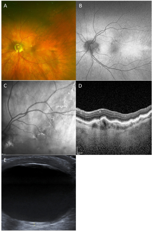
Methods: Demographics and multimodal imaging features of 16 eyes of 13 patients with choroidal macrovessels were reviewed. The multimodal imaging included colour fundus photography, fundus autofluorescence (FAF), spectral domain enhanced depth imaging optical coherence tomography (OCT), en face OCT, OCT-angiography (OCT-A), B-scan ultrasonography (US), fluorescein angiography (FFA) and indocyanine green angiography (ICGA).

Results: Three patients had bilateral involvement. On colour fundus photography, three patterns were evident (a clearly visible orange-red vessel; a track of pigmentary changes; spots of mild pigmentary changes). Vessel orientation was horizontal (11 eyes), oblique (4 eyes) or vertical (1 eye). In 2 eyes, the vessel was extra-macular. OCT in all cases showed a hyporeflective choroidal area with posterior shadowing and elevation of the overlying retina. Subretinal fluid was present in 4 eyes. FAF (12 eyes) was normal (7 eyes) or showed a hypofluorescent/hyperfluorescent track (4 eyes) or linear hyperautofluorescence (1 eye). En-face OCT (2 eyes) revealed the course of the macrovessel at the level of choroid and choriocapillaris. On OCT-A (2 eyes) the vessel had a reflectivity similar to surrounding vessels but larger diameter. B-scan US (8 eyes) showed a nodular hypoechogenic lesion. FFA (5 eyes) showed early focal hyperfluorescence (4 eyes) not increasing in later phases, or was normal (1 eye). ICGA (6 eyes) showed early hyperfluorescence of the vessel.

Conclusions: Choroidal macrovessels can mimic other entities, leading to underdiagnosis. Appreciating relevant features on different imaging modalities will aid a correct diagnosis.

Keywords: choroid; diagnostic tests/investigation; retina.


Conflict of interest statement

Competing interests: None declared.

Figures

Figure 1
Figure 1
Colour fundus photograph, FAF, near-infrared image, EDI-OCT and ICGA of a patient with type I CM. (A) Colour photograph shows a serpiginous lesion extending horizontally from the macula towards the temporal periphery. (B) FAF shows a hypo/hyperautofluorescent pattern in correspondence of the vessel. (C) Near-infrared image showing hyper-reflective and hyporeflective spots in a track-like fashion. (D) EDI-OCT through the macula shows the foveal cm. (E) ICGA shows the early hyperfluorescence and the serpiginous shape of the vessel. (F, G) ICGA in the later phases shows a reduced fluorescence similar to the surrounding vessels with hypofluorescent spots. EDI, enhanced depth imaging; FAF, fundus autofluorescence; ICGA, indocyanine green angiography; OCT, optical coherence tomography.
Figure 2
Figure 2
Pseudocolour fundus image, FAF, near-infrared image, EDI-OCT and B-scan us of a patient with type II CM. (A) Pseudocolour image shows RPE mottling in a track-like fashion. (B) FAF shows a hyperfluorescent and hypoautofluorescent track. (C) Near-infrared image shows hyper-reflectivity and hyporeflectivity. (D) EDI-OCT shows a hollow elevated choroidal area, occupying the entire choroid that is focally thickened. (E) On B-scan US, the CM presents as a nodular lesion with low internal echogenicity. CM, choroidal macrovessel; EDI, enhanced depth imaging; FAF, fundus autofluorescence; OCT, optical coherence tomography; RPE, retinal pigment epithelium; US, ultrasonography.
Figure 3
Figure 3
Composite multicolour, FAF, near-infrared image, EDI-OCT and OCT-A of type III CM temporal to the fovea. (A) Composite multicolour image shows mild RPE changes. (B) FAF shows tiny hyperautofluorescent spots. (C) Near-infrared image shows an area of increased reflectance. (D) EDI-OCT shows choroidal hollowness and mild elevation of the overlying retina. (E) Pseudocolour superimposition of all OCT-A scans highlights two distinguishable purple alterations. (F) structural en face choroid scan showing the CM. (G, H) Scans at the level of choriocapillaris (G) and choroid (H), both showing a vessel having a larger diameter and a hyporeflective centre. CM, choroidal macrovessel; EDI, enhanced depth imaging; FAF, fundus autofluorescence; OCT-A, optical coherence tomography-angiography; RPE, retinal pigment epithelium.

Similar articles

Cited by

References

    1. Lima LH, Laud K, Chang LK, et al. . Choroidal macrovessel. Br J Ophthalmol 2011;95:1333–4. 10.1136/bjo.2009.176800 - DOI - PubMed
    1. Dalvin LA, Pennington JD, Mashayekhi A, et al. . Multimodal imaging of macular choroidal macrovessel: a report of two cases. Retin Cases Brief Rep 2018;0:1–6. 10.1097/ICB.0000000000000776 - DOI - PubMed
    1. Mahroo OA, Puertas RA, Saihan Z. Swept source OCT image of a choroidal Macrovessel. Ophthalmol Retina 2017;1:281. 10.1016/j.oret.2016.12.007 - DOI - PubMed
    1. Casalino G, De Simone L, Scialdone A, et al. . Choroidal macrovessel: optical coherence tomography angiography and imaging features. Indian J Ophthalmol 2019;67:1168–70. 10.4103/ijo.IJO_2085_18 - DOI - PMC - PubMed
    1. Ehlers JP, Rayess H, Spaide RF. Isolated choroidal macrovessel: a tracklike choroidal lesion. Can J Ophthalmol 2014;49:e158–60. 10.1016/j.jcjo.2014.09.006 - DOI - PMC - PubMed

Publication types

MeSH terms