. 2021 Jan 12;118(2):e2022826118.
doi: 10.1073/pnas.2022826118.

Cryoelectron-microscopy structure of the enteropathogenic Escherichia coli type III secretion system EspA filament

Affiliations

Cryoelectron-microscopy structure of the enteropathogenic Escherichia coli type III secretion system EspA filament

Weili Zheng et al. Proc Natl Acad Sci U S A. .

Abstract

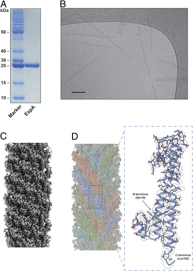
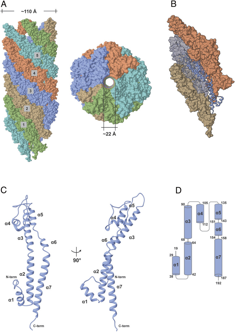
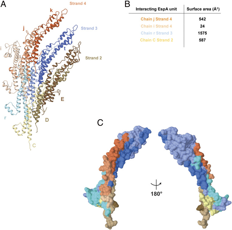
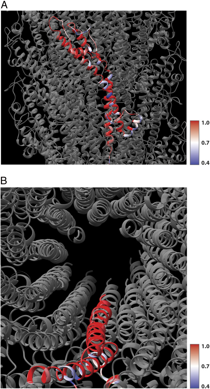
Enteropathogenic Escherichia coli (EPEC) and enterohemorrhagic Escherichia coli (EHEC) utilize a macromolecular type III secretion system (T3SS) to inject effector proteins into eukaryotic cells. This apparatus spans the inner and outer bacterial membranes and includes a helical needle protruding into the extracellular space. Thus far observed only in EPEC and EHEC and not found in other pathogenic Gram-negative bacteria that have a T3SS is an additional helical filament made by the EspA protein that forms a long extension to the needle, mediating both attachment to eukaryotic cells and transport of effector proteins through the intestinal mucus layer. Here, we present the structure of the EspA filament from EPEC at 3.4 Å resolution. The structure reveals that the EspA filament is a right-handed 1-start helical assembly with a conserved lumen architecture with respect to the needle to ensure the seamless transport of unfolded cargos en route to the target cell. This functional conservation is despite the fact that there is little apparent overall conservation at the level of sequence or structure with the needle. We also unveil the molecular details of the immunodominant EspA epitope that can now be exploited for the rational design of epitope display systems.

Keywords: attaching/effacing pathogens; bacterial pathogenesis; intestinal epithelium.


Conflict of interest statement

The authors declare no competing interest.

Figures

Fig. 1.
Fig. 1.
Purification of the EspA filament and cryo-EM reconstruction at 3.4 Å. (A) SDS-PAGE of purified EspA filament. (B) Electron micrograph of EspA pili. (Scale bar, 50 nm.) (C) Side view of the EspA filament cryo-EM map. (D) The EspA filament cryo-EM map was fitted with the derived atomic model (Left), and the five strands are colored in cyan, orange, blue, brown, and green. Details of the EspA subunit density (Right) with the EspA model built into the density from Ser19 to Lys192 and shown in ribbon representation.
Fig. 2.
Fig. 2.
Overall architecture of the EspA filament and the EspA subunit. (A) Surface representations of side (Left) and top (Right) views of the EspA filament structure. Each of the five strands is shown in a different color. (B) Localization of an individual subunit within the EspA filament strand 3. (C) Ribbon representation of the EspA protein structure with the N and C terminus and all the secondary structures labeled. (D) Topology secondary structure diagram of the EspA protein with α-helices represented as cylinders and showing the first and last residues in each helix.
Fig. 3.
Fig. 3.
EspA–EspA interaction network in the filament. (A) Each EspA subunit interacts with eight other subunit chains within the filament. The reference chain s strand 3 (blue) interacts with the neighboring chains r (light blue) and t (dark blue) from the same strand 3, with chains i (light orange), j (orange), and k (dark orange) from strand 4 and with chains C (light brown), D (brown), and E (dark brown) from strand 2. (B) Buried surface area of the EspA–EspA interfaces using the same chain reference and the color coding as in A. The other four interfaces made with the reference chain s (with chains k, t, D, and E) are symmetry-related to these four. (C) Mapping of the EspA–EspA interactions in the reference subunit (chain s strand 3, blue color). The interactions made by the eight neighboring subunits are mapped in the reference subunit using the same color code as in A.
Fig. 4.
Fig. 4.
The EspA filament and the T3SS needle form a seamless conduit. (A) Cryo-EM maps of the T3SS apparatus (EMD-8913 and EMD-8914) and T3SS needle (EMD-8924), with EspA filament map located on top of the PgrI T3SS needle (Left). Central cross-section of the same composite map (Right). (B) Surface representation of lumen segments from the EspA filament (dark green) and the T3SS needle (gray). The volumes were determined using a fixed surface height of 60 Å in both calculations (Left). Surface representation of the residues exposed in the EspA filament lumen (Arg183, Ser189, and Lys192) and PrgI needle lumen (Lys66, Gln77, Asp70, Asn78, and Arg80) highlighting the right-handed spiral interior in both filaments. (C) Distribution of residues charges within the EspA filament, PrgI T3SS needle (PDB:6DWB), and C. jejuni flagellar filament (PDB: 6X80).
Fig. 5.
Fig. 5.
The conservation of residues in other EspA proteins. (A) Four other EspA proteins (having from 79 to 89% overall sequence identity with EPEC EspA) have been aligned, and the conservation at every residue is mapped onto a single EspA subunit in the filament, with red indicating 100% sequence identity at that position and blue indicating that 40% of the sequences have the same residue at this position. Almost all of the nonconserved residues are on the outer surface of the filament. (B) In contrast, a top view shows that all of the residues that line the lumen are highly conserved in these homologs.

Similar articles

Cited by

References

    1. Chen H. D., Frankel G., Enteropathogenic Escherichia coli: Unravelling pathogenesis. FEMS Microbiol. Rev. 29, 83–98 (2005). - PubMed
    1. Pennington H., Escherichia coli O157. Lancet 376, 1428–1435 (2010). - PubMed
    1. Garmendia J., Frankel G., Crepin V. F., Enteropathogenic and enterohemorrhagic Escherichia coli infections: Translocation, translocation, translocation. Infect. Immun. 73, 2573–2585 (2005). - PMC - PubMed
    1. Deng W., et al. , Assembly, structure, function and regulation of type III secretion systems. Nat. Rev. Microbiol. 15, 323–337 (2017). - PubMed
    1. Wilson R. K., Shaw R. K., Daniell S., Knutton S., Frankel G., Role of EscF, a putative needle complex protein, in the type III protein translocation system of enteropathogenic Escherichia coli. Cell. Microbiol. 3, 753–762 (2001). - PubMed

Publication types

MeSH terms

Substances

LinkOut - more resources