Review
. 2021 Jan 15;206(2):264-272.
doi: 10.4049/jimmunol.2001060.

Afferent Lymphatic Transport and Peripheral Tissue Immunity

Affiliations
Review

Afferent Lymphatic Transport and Peripheral Tissue Immunity

Maria M Steele et al. J Immunol. .

Abstract

Lymphatic vessels provide an anatomical framework for immune surveillance and adaptive immune responses. Although appreciated as the route for Ag and dendritic cell transport, peripheral lymphatic vessels are often not considered active players in immune surveillance. Lymphatic vessels, however, integrate contextual cues that directly regulate transport, including changes in intrinsic pumping and capillary remodeling, and express a dynamic repertoire of inflammatory chemokines and adhesion molecules that facilitates leukocyte egress out of inflamed tissue. These mechanisms together contribute to the course of peripheral tissue immunity. In this review, we focus on context-dependent mechanisms that regulate fluid and cellular transport out of peripheral nonlymphoid tissues to provide a framework for understanding the effects of afferent lymphatic transport on immune surveillance, peripheral tissue inflammation, and adaptive immunity.


Figures

Figure 1.
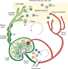
Figure 1.. The lymphatic anatomy of immune surveillance.
Under basal conditions, immune surveillance is regulated by constitutive lymph transport and the trafficking of lymphocytes (naive, memory and regulatory) and antigen presenting cells between peripheral tissue, lymphoid, and systemic compartments. Vascular transudate generates directional interstitial fluid flows in the direction of draining, low pressure lymphatic capillaries, where open, discontinuous button-like junctions (dotted red lines) facilitate passive uptake and permit integrin-independent cell transmigration. Oncotic gradients and interstitial fluid flow (0.1–1 μm/s) enrich afferent lymph for tissue-derived factors, antigens, large particulates, and protein complexes that reflect the immunological status of the tissue from which it drains. Collecting lymphatic vessels propel fluid towards draining lymph nodes through intrinsic pumping mechanisms regulated by invested LMCs, and continuous zipper-like intercellular junctions (solid red lines) restrict vessel permeability. Leukocytes migrating through afferent lymph are predominantly of memory and regulatory phenotypes, distinct from both efferent lymph and blood where naive T cells also recirculate in search of cognate antigen loaded on migratory DCs and other antigen presenting cells. Resident memory T cells are defined by their exclusion from afferent lymph and long-term retention in peripheral, non-lymphoid tissue at basal conditions. Dendritic cells (DC); naive T cells (TN); memory T cells (TMem); resident memory T cells (TRM); regulatory T cells (TREG); lymphatic muscle cell (LMC).
Figure 2.
Figure 2.. T cell egress from peripheral tissues via lymphatic capillaries.
Peripheral lymphatic capillaries mediate the directional homing and transmigration of T lymphocytes from steady state and inflamed peripheral tissues. (1) Under basal conditions, the trafficking of memory and regulatory lymphocytes is largely dependent on the homeostatic chemokine, CCL21, constitutively expressed by peripheral lymphatic capillaries. (2) Inflammatory cytokines, such as TNFɑ and IL-1β, activate regional blood and lymphatic vessels, increasing vascular permeability, interstitial fluid flow, and inducing lymphatic endothelial production of a diverse repertoire of chemokines and adhesion molecules that presumably direct the egress of T cells. Activated CD4+ and CD8+ effector T cells are recruited to inflamed peripheral tissues where recognition of cognate antigen, downregulates lymphatic homing receptors (S1PRs and CCRs), supports tissue retention, and ultimately long-term residence as TRM. IFNγ produced by antigen-specific CD8+ T cell responses further activates the lymphatic vasculature to induce expression of MHCII and PD-L1, which may act to negatively regulate ongoing cytotoxic immunity. Active mechanisms increase the overall abundance of lymph-borne lymphocytes. (3) S1P acts both on CD4+ T cells, which use S1P1 and S1P4 to enter afferent lymph, and lymphatic endothelial cells where S1P2 regulates VCAM expression and transendothelial migration. Additionally, during acute inflammatory processes in skin, CCR7 and ICAM-1 is required for T cell egress via lymphatic vessels, while CCR7-dependence is lost in chronic inflammation. (4) In grafts, TREG cell migration to lymph nodes is necessary to limit alloimmune responses and transmigrating TREG condition the lymphatic endothelium to express higher levels of VCAM through activation of LTβR that supports subsequent egress of inflammatory lymphocytes (CD4+ and CD8+ T cells) to resolve peripheral tissue inflammation. (5) Lymphatic capillaries also directly regulate peripheral tissue inflammation by scavenging inflammatory chemokines through expression of decoy receptors (ACKR2/D6) limiting their effect in peripheral tissue and transport to lymph nodes. Memory T cells (TMem); effector T cells (TEff); resident memory T cells (TRM); regulatory T cells (TREG); antigen presenting cell (APC).

References

    1. Oliver G, Kipnis J, Randolph GJ, and Harvey NL. 2020. The Lymphatic Vasculature in the 21st Century: Novel Functional Roles in Homeostasis and Disease. Cell 182: 270–296. - PMC - PubMed
    1. Petrova TV, and Koh GY. 2020. Biological functions of lymphatic vessels. Science 369: eaax4063. - PubMed
    1. Santambrogio L 2018. Lymph Formation and Composition. Lymphedema 139–152.
    1. Baluk P, Fuxe J, Hashizume H, Romano T, Lashnits E, Butz S, Vestweber D, Corada M, Molendini C, Dejana E, and McDonald DM. 2007. Functionally specialized junctions between endothelial cells of lymphatic vessels. J Exp Med 204: 2349–2362. - PMC - PubMed
    1. Pflicke H, and Sixt M. 2009. Preformed portals facilitate dendritic cell entry into afferent lymphatic vessels. J Exp Med 206: 2925–2935. - PMC - PubMed

Publication types

LinkOut - more resources