. 2021 Jan 4;12(1):93.
doi: 10.1038/s41467-020-20310-0.

The role of water in host-guest interaction

Affiliations

The role of water in host-guest interaction

Valerio Rizzi et al. Nat Commun. .

Abstract

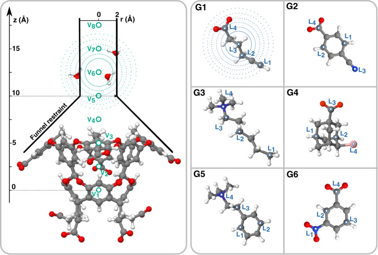
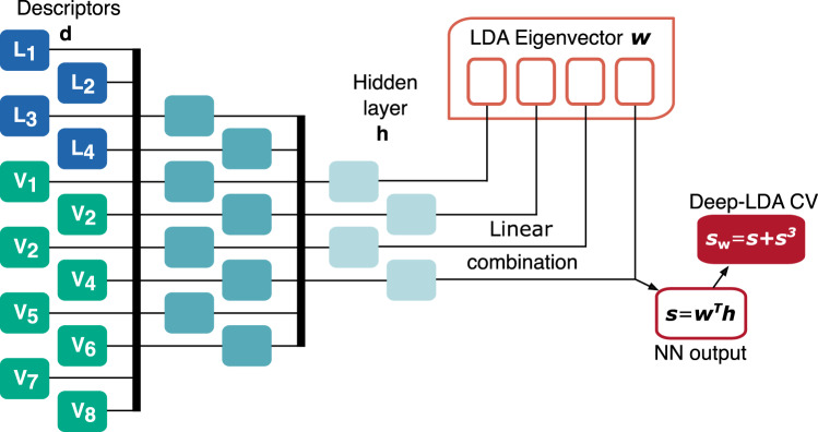
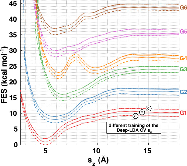
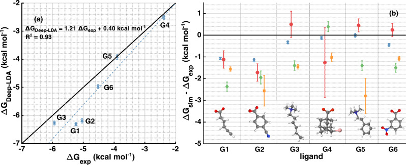
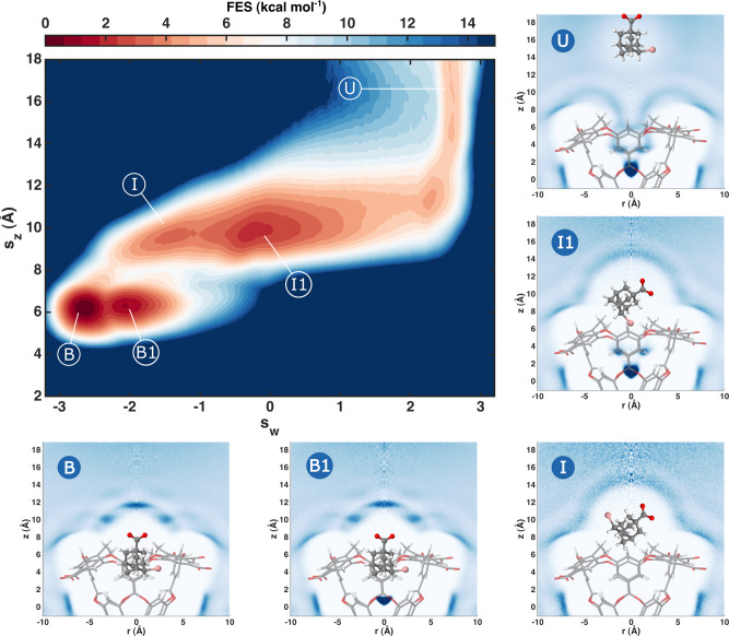
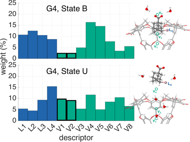
One of the main applications of atomistic computer simulations is the calculation of ligand binding free energies. The accuracy of these calculations depends on the force field quality and on the thoroughness of configuration sampling. Sampling is an obstacle in simulations due to the frequent appearance of kinetic bottlenecks in the free energy landscape. Very often this difficulty is circumvented by enhanced sampling techniques. Typically, these techniques depend on the introduction of appropriate collective variables that are meant to capture the system's degrees of freedom. In ligand binding, water has long been known to play a key role, but its complex behaviour has proven difficult to fully capture. In this paper we combine machine learning with physical intuition to build a non-local and highly efficient water-describing collective variable. We use it to study a set of host-guest systems from the SAMPL5 challenge. We obtain highly accurate binding free energies and good agreement with experiments. The role of water during the binding process is then analysed in some detail.


Conflict of interest statement

The authors declare no competing interests.

Figures

Fig. 1
Fig. 1. Sketch of the octa-acid host OAMe with the funnel restraint geometry and the guest molecules from the SAMPL5 challenge.
We indicate the position of the points where the descriptors are centred and hint at their spatial outreach by drawing surfaces at a constant radius around some of them.
Fig. 2
Fig. 2. Schematics of the Deep-LDA architecture used in this work.
The descriptors d are fed to a NN that generates s as a linear combination of the last NN hidden layer h and the LDA eigenvector w. The Deep-LDA CV is then sw = s + s3.
Fig. 3
Fig. 3. Free energy surfaces projected along the host–guest distance.
For each of the six ligands, we compute the free energy along the sz variable using a standard umbrella-sampling-like reweighting formula to recover the unbiased distribution. The shaded areas indicate the errors, whose calculation is detailed in the Supplementary Methods. To ensure that the results do not depend on a specific realisation of the Deep-LDA CV, we repeat the training three times by using different initial weights of the NN. The resulting CVs are denoted as swa, swb, swc, and the corresponding FES are indicated, respectively, by dashed, dotted, and dash-dotted lines. For clarity, curves related to the same ligand but with different CVs are shifted by 1 kcal mol−1, while the shift between different ligand curves is 5 kcal mol−1.
Fig. 4
Fig. 4. Comparison of the binding free energies with experiments and other calculations.
In a, we plot the value of ΔG obtained from the Deep-LDA simulations (in blue crosses) for every ligand versus the experimental values and show the corresponding linear fit. In b, we report their difference with the experimental values and compare them with other computational results performed using the same simulation setup. Results from ref. are indicated with red circles, from ref. in green diamonds, and from ref. in yellow squares.
Fig. 5
Fig. 5. Binding FES of ligand G4 with a study of the water presence in the visited states.
We show the two-dimensional FES of the ligand G4 with respect to sz and Deep-LDA CV sw. Different adjacent colours correspond to a free energy difference of 1 kBT ≈ 0.6 kcal mol−1. We highlight some relevant states over which we perform plain molecular dynamics (MD) simulations to measure the presence of water. We show histograms of the water oxygen atoms’ density in cylindrical coordinates zr. Each histogram is normalised by the density value in its top-right corner and darker colours correspond to higher-water-density regions. The position of the ligand in these plots is illustrative.
Fig. 6
Fig. 6. Descriptors’ relative weights for guest G4.
Following the derivative-based ranking from ref. , we show the relative weight that each descriptor has in the Deep-LDA CV in the bound and unbound states of guest G4. We show the average weight over the three different Deep-LDA CVs that we trained. The V1, V2 descriptors, which measure the number of molecules inside the pocket, are outlined to mark the significant change in their contribution between the two states.

References

    1. Michel J, Essex JW. Prediction of protein-ligand binding affinity by free energy simulations: assumptions, pitfalls and expectations. J. Computer-Aided Mol. Des. 2010;24:639–658. doi: 10.1007/s10822-010-9363-3. - DOI - PubMed
    1. Mobley DL, Gilson MK. Predicting binding free energies: frontiers and benchmarks. Annu. Rev. Biophys. 2017;46:531–558. doi: 10.1146/annurev-biophys-070816-033654. - DOI - PMC - PubMed
    1. Bronowska, A. In Thermodynamics - Interaction Studies - Solids, Liquids and Gases Vol. i (InTech, 2011) https://www.intechopen.com/books/thermodynamics-interaction-studies-soli....
    1. Limongelli V, Bonomi M, Parrinello M. Funnel metadynamics as accurate binding free-energy method. Proc. Natl Acad. Sci. USA. 2013;110:6358–6363. doi: 10.1073/pnas.1303186110. - DOI - PMC - PubMed
    1. Tiwary P, Mondal J, Berne BJ. How and when does an anticancer drug leave its binding site? Sci. Adv. 2017;3:e1700014. doi: 10.1126/sciadv.1700014. - DOI - PMC - PubMed

Publication types

LinkOut - more resources