. 2021 Jan 4;7(1):2.
doi: 10.1038/s41531-020-00147-6.

Plasma-borne indicators of inflammasome activity in Parkinson's disease patients

Affiliations

Plasma-borne indicators of inflammasome activity in Parkinson's disease patients

Faith L Anderson et al. NPJ Parkinsons Dis. .

Abstract

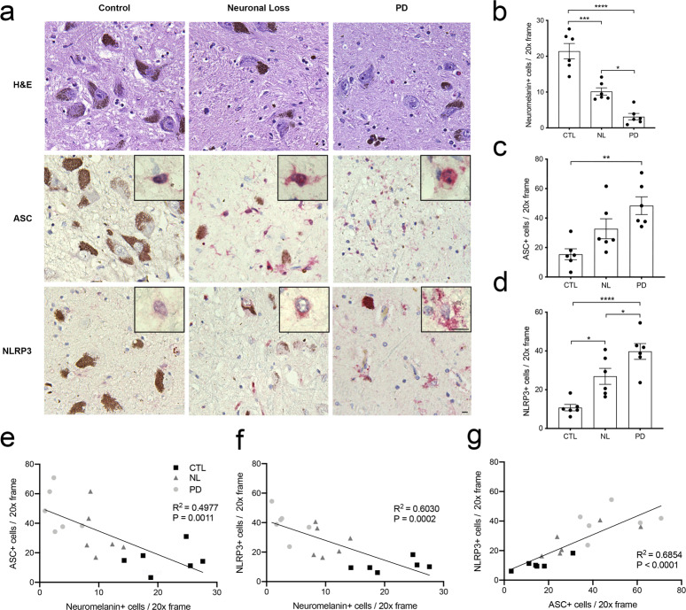
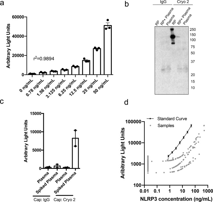
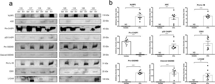
Parkinson's disease (PD) is a neurodegenerative disorder characterized by motor and non-motor symptoms and loss of dopaminergic neurons of the substantia nigra. Inflammation and cell death are recognized aspects of PD suggesting that strategies to monitor and modify these processes may improve the management of the disease. Inflammasomes are pro-inflammatory intracellular pattern recognition complexes that couple these processes. The NLRP3 inflammasome responds to sterile triggers to initiate pro-inflammatory processes characterized by maturation of inflammatory cytokines, cytoplasmic membrane pore formation, vesicular shedding, and if unresolved, pyroptotic cell death. Histologic analysis of tissues from PD patients and individuals with nigral cell loss but no diagnosis of PD identified elevated expression of inflammasome-related proteins and activation-related "speck" formation in degenerating mesencephalic tissues compared with controls. Based on previous reports of circulating inflammasome proteins in patients suffering from heritable syndromes caused by hyper-activation of the NLRP3 inflammasome, we evaluated PD patient plasma for evidence of inflammasome activity. Multiple circulating inflammasome proteins were detected almost exclusively in extracellular vesicles indicative of ongoing inflammasome activation and pyroptosis. Analysis of plasma obtained from a multi-center cohort identified elevated plasma-borne NLRP3 associated with PD status. Our findings are consistent with others indicating inflammasome activity in neurodegenerative disorders. Findings suggest mesencephalic inflammasome protein expression as a histopathologic marker of early-stage nigral degeneration and suggest plasma-borne inflammasome-related proteins as a potentially useful class of biomarkers for patient stratification and the detection and monitoring of inflammation in PD.


Conflict of interest statement

The authors declare no competing interests.

Figures

Fig. 1
Fig. 1. Elevated ASC and NLRP3 protein expression observed during multiple stages of mesencephalic neurodegeneration.
a Post-mortem mesencephalic tissues were obtained from healthy control subjects (CTL) (n = 6), asymptomatic subjects with loss of SNpC neurons (NL) (n = 6), and clinically confirmed PD patients (PD) (n = 6). Tissues were sectioned at 5 µM and stained with H&E (top row), anti-ASC (middle row, red), or anti-NLRP3 antibodies (bottom row, red). Scale bars in 20× field and 60× inset represent 10 μm. b Fewer neuromelanin-positive neurons were observed in the SNpC in both NL tissues and PD tissues relative to healthy controls (****P < 0.0001, ***P = 0.0002, *P = 0.0111). c More ASC-positive cells were observed in the PD tissues relative to controls (**P = 0.0024). d More NLRP3-positive cells were observed in the NL tissues relative to CTL tissues (*P = 0.0132), while there were significantly more NLRP3-positive cells observed in the PD tissues relative to NL tissues (*P = 0.0487) and CTL tissues (****P < 0.0001). All quantification was performed by counting 10 distinct fields per section. P-values were determined using one-way ANOVA with multiple comparisons, error bars in bd represent s.e.m. e ASC-immunoreactive cells compared to neuromelanin-positive cells per tissue in three histologic groups (R2 = 0.4977, P = 0.0011). f NLRP3-immunoreactive cells were statistically associated with reduced number of neuromelanin-positive cells across three histologic groups (R2 = 0.6030, P = 0.0002). g Correlation between number of ASC-immunoreactive and NLRP3-immunoreactive cells (R2 = 0.6854, P < 0.0001).
Fig. 2
Fig. 2. Development of an electrochemiluminescent-based assay for the detection of inflammasome-related protein NLRP3.
Anti-NLRP3 antibodies were tested in different capture and detection antibody pairing orientations in 96-well electrode-containing immunosorbent plates (Meso Scale Diagnostics (MSD), Rockville, MD) for their ability to detect NLRP3 recombinant protein. Once an antibody pair was found, the capture antibody was biotinylated and assay conditions were optimized using streptavidin-coated 96-well electrode-containing immunosorbent plates. a The antibody pair identified was able to detect recombinant NLRP3 protein in 1% BSA (r2 = 0.9894) down to a concentration of 0.78 ng/mL. R2 was calculated using linear regression. b NLRP3 detection was confirmed through immunoprecipitation (IP), where the capture antibody was used as the pull-down antibody and the detection antibody was used as the immunoblotting antibody. c Clean plasma or plasma spiked with 12.5 ng/mL of recombinant NLRP3 protein was run on the optimized electrochemiluminescent assay using biotinylated mouse IgG or biotinylated Cryo 2 as the capture antibody in order to ensure antibody-pair specificity and low non-specific binding. d NLRP3 protein concentration was plotted against electrochemiluminescent read-out (Arbitrary Light Units) for the representative standard curve and all samples analyzed. Axes are on the logarithmic scale. Samples classified as “non-detectable” were imputed by taking the minimum positive value divided by 2. Error bars in a and c represent s.e.m.
Fig. 3
Fig. 3. Inflammasome-related proteins are present in extracellular vesicles isolated from human plasma samples.
a Extracellular vesicles (EVs) were isolated from fresh plasma samples using the ExoQuick Plasma Prep with Thrombin (EXOQ5TM-1, System Biosciences, Palo Alto, CA). The EV fractions were lysed and sonicated before analysis. The soluble and EV fractions from 8 plasma samples were analyzed via SDS–PAGE and subsequent immunoblotting. Anti-NLRP3, anti-ASC, anti-CASP1, anti-GSDMD, and anti-IL-1B antibodies were used for the detection of inflammasome-related proteins. Anti-CD81 and anti-L1CAM antibodies were used to confirm the purity of the EV fraction and assess the presence of neural-derived EVs, respectively. b The presence of inflammasome-related proteins was most often found solely in the EV fraction and the detection of EV-specific markers was found only in the EV fraction. All statistical analyses were performed using unpaired Students t-tests (NLRP3, ****P < 0.0001) (ASC, ***P = 0.0002) (Pro-CASP1, ****P < 0.0001) (p20 CASP1, ****P < 0.0001) (Pro-GSDMD, ****P < 0.0001) (Cleaved GSDMD, ****P < 0.0001) (Pro IL-1B, ****P < 0.0001) (CD81, ****P < 0.0001) (L1CAM, **P = 0.0018). Error bars represent s.e.m.

References

    1. Kalia LV, Lang AE. Parkinson’s disease. Lancet. 2015;386:896–912. doi: 10.1016/S0140-6736(14)61393-3. - DOI - PubMed
    1. Poewe W, et al. Parkinson disease. Nat. Rev. Dis. Prim. 2017;3:17013. doi: 10.1038/nrdp.2017.13. - DOI - PubMed
    1. Del Rey NL, et al. Advances in Parkinson’s disease: 200 years later. Front. Neuroanat. 2018;12:113. - PMC - PubMed
    1. Kulisevsky J, Oliveira L, Fox SH. Update in therapeutic strategies for Parkinson’s disease. Curr. Opin. Neurol. 2018;31:439–447. doi: 10.1097/WCO.0000000000000579. - DOI - PubMed
    1. Hirsch EC, et al. The role of glial reaction and inflammation in Parkinson’s disease. Ann. N. Y. Acad. Sci. 2003;991:214–228. doi: 10.1111/j.1749-6632.2003.tb07478.x. - DOI - PubMed