. 2021 May;28(5):1548-1562.
doi: 10.1038/s41418-020-00685-9. Epub 2021 Jan 4.

Loss of ferroportin induces memory impairment by promoting ferroptosis in Alzheimer's disease

Affiliations

Loss of ferroportin induces memory impairment by promoting ferroptosis in Alzheimer's disease

Wen-Dai Bao et al. Cell Death Differ. 2021 May.

Erratum in

Abstract

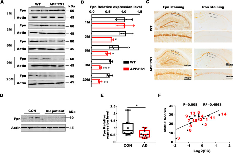
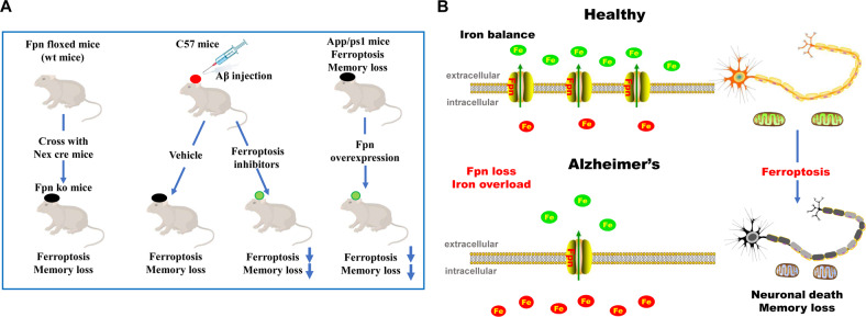
Iron homeostasis disturbance has been implicated in Alzheimer's disease (AD), and excess iron exacerbates oxidative damage and cognitive defects. Ferroptosis is a nonapoptotic form of cell death dependent upon intracellular iron. However, the involvement of ferroptosis in the pathogenesis of AD remains elusive. Here, we report that ferroportin1 (Fpn), the only identified mammalian nonheme iron exporter, was downregulated in the brains of APPswe/PS1dE9 mice as an Alzheimer's mouse model and Alzheimer's patients. Genetic deletion of Fpn in principal neurons of the neocortex and hippocampus by breeding Fpnfl/fl mice with NEX-Cre mice led to AD-like hippocampal atrophy and memory deficits. Interestingly, the canonical morphological and molecular characteristics of ferroptosis were observed in both Fpnfl/fl/NEXcre and AD mice. Gene set enrichment analysis (GSEA) of ferroptosis-related RNA-seq data showed that the differentially expressed genes were highly enriched in gene sets associated with AD. Furthermore, administration of specific inhibitors of ferroptosis effectively reduced the neuronal death and memory impairments induced by Aβ aggregation in vitro and in vivo. In addition, restoring Fpn ameliorated ferroptosis and memory impairment in APPswe/PS1dE9 mice. Our study demonstrates the critical role of Fpn and ferroptosis in the progression of AD, thus provides promising therapeutic approaches for this disease.


Conflict of interest statement

The authors have declared that no conflicts of interest exist.

Figures

Fig. 1
Fig. 1. Fpn is downregulated in the hippocampus of AD mice and patient brain tissues.
A Representive protein level of Fpn in the hippocampus of APPswe/PS1dE9 (APP/PS1) mice at different ages (M: month) and the age-matched wild-type littermates (WT). B The quantification for protein level of Fpn in the hippocampus of APPswe/PS1dE9 (APP/PS1) mice (n = 3). C Immuno-histochemistry of Fpn and the DAB-enhanced Perl’s Prussian blue iron staining in the hippocampus of APPswe/PS1dE9 (APP/PS1) mice and the age-matched wild-type littermates (WT) at 9 months old. D The protein level of Fpn in brain tissues of AD patients and control (CON) sample (frontal cortex, 3 con vs 4 AD). E The fold change of the quantification for the protein level of Fpn in cortical brain tissues of AD patients (n = 10) compared to corresponding control (CON) samples (n = 9). F Correlation analysis between the Fpn protein level (Log2FC: The log2 fold-change of the Fpn protein expression level compared to the average of the controls) and the MMSE scores of subjects. The corresponding serial numbers of the human samples are marked with red number adjacent to each point (n = 14). Protein expression levels were detected by western blotting. Data are shown as the mean ± SD of at least three independent experiments. Statistical analyses were carried out using two-way ANOVA and mutiple t-test. *p < 0.05; **p < 0.01; ***p < 0.001. Detailed Statistical analyses are included with the Supplementary Table S4.
Fig. 2
Fig. 2. Fpnfl/fl/NEXcre mice developed brain atrophy.
A The protein level of the Fpn in the primary neurons hippocampus of Fpnfl/fl/NEXcre mice. B The representative images of the whole brain of Fpnfl/fl/NEXcre mice and age-matched floxed littermates (Fpnfl/fl). C The weight of the hippocampus and whole brain in Fpnfl/fl/NEXcre mice (n = 5) and age-matched controls (Fpnfl/fl) (n = 5) littermates at 1 month and 3 month of age. D The representative MRI images (left) and relative quantitative data (right) from the brains of Fpnfl/fl/NEXcre mice and age-matched wild type littermates at 1 month old (n = 3–4). E Nissl staining of Fpnfl/fl/NEXcre mice at 3 month of age (Right panels are the magnification image as indicated in the left panels). F Quantitative fold change of the number of neurons in hippocampus by Nissl staining of Fpnfl/fl/NEXcre mice (n = 5) and age-matched wild type littermates (n = 5) at 3 months old. G DAB-enhanced Perl’s Prussian blue iron staining of Fpnfl/fl/NEXcre mice brains at 3 months old (right panels are the magnified images as indicated in the left panels). H The tissue iron content (μg/g) in the hippocampus of Fpnfl/fl/NEXcre mice at 1 month old and 3 months old (n = 4). Protein expression levels were detected by western blotting. Data are shown as the mean ± SD of at least three independent experiments. Statistical analyses were carried out using two-way ANOVA and multiple t-tests. *p < 0.05; **p < 0.01; ***p < 0.001. Detailed Statistical analyses are included with the Supplementary Table S4.
Fig. 3
Fig. 3. Fpnfl/fl/NEXcre mice developed cognitive impairment.
A The static time (Static T), moved time (Move T), moved distance (Move D) and the time spent at the center (Center T), corner (Corner T), side (Side T) of Fpnfl/fl/NEXcre mice (n = 6) and age-matched wild type (WT) littermates (n = 6) in an open field test. B The representative searching trace (left) and the latency in the learning stages in the Morris water maze of Fpnfl/fl/NEXcre mice (n = 18) and age-matched floxed littermates at 10–12 months old (n = 20). C The moved distance of Fpn ko mice (n = 18) and littermate control (n = 20) in Morris water maze. D The time in the target quadrant and (E) as well as the latency to reach a hidden platform on day 7 of the ko mice. F Decreased time spent freezing in the contextual fear conditioning in Fpnfl/fl/NEXcre mice (Fpnfl/fl, n = 20, Fpnfl/fl/NEXcre, n = 18). G The percentage time spent freezing during training or before tone in the fear conditioning test of these mice. H The Fpn protein level in hippocampus of C57 mice injected with lentivirus expressing shRNA against fpn (FPN-RNAi) or scrambled hairpin (Con virus). I The representative searching trace (left) and the latency in the learning stages (right) in the Morris Water Maze test of C57 mice injected with lentivirus expressing shRNA against fpn (FPN-RNAi) (n = 10) or scrambled hairpin (Con virus) (n = 8). J The moved distance of these mice in Morris water maze tests. K The time spent in the target quadrant and (L) the latency to reach a hidden platform on day 7 of these mice. Data are shown as the mean ± SD of at least three independent experiments. Statistical analyses were carried out using two-way ANOVA and mutiple t-tests. *p < 0.05; **p < 0.01; ***p < 0.001. Detailed Statistical analyses are included with the Supplementary Table 4.
Fig. 4
Fig. 4. Both Fpnfl/fl/NEXcre and AD mice developed features of ferroptosis.
A Transmission electron microscopy pictures of perinuclear area of hippocampal neurons from Fpnfl/fl, Fpnfl/fl/NEXcre, WT and APPswe/PS1dE9 mice at 9 months old. WT were age-matched wild type littermates of the transgenic mice. B Mitochondrial area frequency in perinuclear compartment of these mice. Calculated from n > 100 mitochondria from n > 10 pictures of 3 mice per group. C The MDA content and D the GSH content in the hippocampus of Fpnfl/fl, Fpnfl/fl/NEXcre, WT and APPswe/PS1dE9 mice at 9 months old. WT were age-matched wild type littermates of the transgenic mice (n = 5). E The protein levels of Gpx-4 were detected in the hippocampus of the WT and APPswe/PS1dE9 mice and F Fpnfl/fl, Fpnfl/fl/NEXcre mice. β-Actin was served as a loading control. G The mRNA level of ACSF, IREB2, CS, RPL8, ATP5G3 and PTGS2 were detected in the hippocampus of the WT (n = 5) and APPswe/PS1dE9 mice (n = 5) and H Fpnfl/fl (n = 5), Fpnfl/fl/NEXcre mice (n = 5). β-Actin was used as an internal control, and results are shown as fold change of the control. Data are shown as the mean ± SD of at least three independent experiments. Statistical analyses were carried out using two-way ANOVA and mutiple t-tests. *p < 0.05; **p < 0.01; ***p < 0.001. Detailed Statistical analyses are included with the Supplementary Table S4.
Fig. 5
Fig. 5. Inhibitors of ferroptosis ameliorated the neuronal death and memory impairment induced by Aβ1–42 aggregation in vitro and in vivo.
A Primary neurons were exposed to 10 μm/20 μm Aβ1–42 and ferroptotic inhibitors for 24 h. The cell viability was accessed by CCK8 assays (n = 4). B After treatment (10 μm Aβ1–42, 100 nM Lip-1 or 1 μm Fer-1), the neurons were stained with PI and DAPI. Representative images and C percentage of PI + /DAPI + cells are shown (n = 10). D Protein levels of Fpn, FTH, and Gpx4 in the hippocampus exposed to Aβ1–42. E Representative images of Nissl and PI staining of the dentate gyrus of the mice exposed to Aβ1–42 and ferroptotic inhibitors. F The quantification for E (vehicle n = 10; Aβ n = 10; Aβ + Lip-1 n = 8; Aβ + Fer-1 n = 8). G The latency during the learning stages (left) and the representative searching trace (right) in the Morris water maze of the mice exposed to Aβ1–42 injection and ferroptotic inhibitors (vehicle, n = 10; Aβ n = 10; Aβ + Fer-1 n = 8; Aβ + Lip-1 n = 8). H The latency to reach a hidden platform as well as (I) time in the target quadrant on day 7 of these mice. Data are shown as the mean ± SD of at least three independent experiments. Statistical analyses were carried out using two-way ANOVA and mutiple t-tests. *p < 0.05; **p < 0.01; ***p < 0.001. Detailed Statistical analyses are included with the Supplementary Table S4.
Fig. 6
Fig. 6. Restoring of Fpn in hippocampus ameliorated ferroptosis and memory loss in APPswe/PS1dE9 mice.
A The latency (left) and the representative searching trace (right) to the platform of APPswe/PS1dE9 mice with AAVs overexpressed full length murine Fpn (Fpn-AAV, n = 10) or corresponding con-AAVs (con AAV, n = 10) injection during the training process at 12 months old. B The time in the target quadrant as well as C the latency to reach a hidden platform on day 7 of APPswe/PS1dE9 mice with con-AAV or Fpn-AAV injection. D Increased time spent freezing in the contextual fear conditioning of the APPswe/PS1dE9 mice with Fpn-AAV injection (n = 10). E The DAB-enhanced Perl’s Prussian blue iron staining of the hippocampus of the AAV injected APPswe/PS1dE9 mice. F The protein levels of Gpx-4 were detected in hippocampus of the APPswe/PS1dE9 mice with con-AAV or Fpn-AAV injection. β-Actin served as a loading control. G The mRNA level of ACSF2, IREB2, CS, RPL8, ATPG53 and PTGS2 were detected in the hippocampus of the APPswe/PS1dE9 mice with con-AAV (n = 5) or Fpn-AAV(n = 5) injection. β-Actin was used as an internal control, and results are shown as fold change of the control. H The MDA content and I the GSH content in the hippocampus of the APPswe/PS1dE9 mice with con-AAV (n = 5) or Fpn-AAV(n = 5) injection compared to the wild-type controls. Data are shown as the mean ± SD of at least three independent experiments. Statistical analyses were carried out using two-way ANOVA and mutiple t-tests. *p < 0.05; **p < 0.01; ***p < 0.001. Detailed Statistical analyses are included with the Supplementary Table S4.
Fig. 7
Fig. 7. Graphical abstract of memory impairment and ferroptosis induced by Fpn loss in Alzheimer’s disease.
A A schematic diagram for the mouse models in this study. B Graphical abstract of the role of ferroptosis induced by deficiency of Fpn in Alzheimer’s disease.

References

    1. Ward R, Zucca FA, Duyn JH, Crichton RR, Zecca L. The role of iron in brain ageing and neurodegenerative disorders. Lancet Neurol. 2014;13:1045–60. doi: 10.1016/S1474-4422(14)70117-6. - DOI - PMC - PubMed
    1. Holcomb L, Gordon MN, McGowan E, Yu X, Benkovic S, Jantzen P, et al. Accelerated Alzheimer-type phenotype in transgenic mice carrying both mutant amyloid precursor protein and presenilin 1 transgenes. Nat Med. 1998;4:97–100. doi: 10.1038/nm0198-097. - DOI - PubMed
    1. Schroder N, Figueiredo LS, de Lima MNM. Role of brain iron accumulation in cognitive dysfunction: evidence from animal models and human studies. J Alzheimers Dis. 2013;34:797–812. doi: 10.3233/JAD-121996. - DOI - PubMed
    1. Roberts BR, Ryan TM, Bush AI, Masters CL, Duce JA. The role of metallobiology and amyloid-beta peptides in Alzheimer’s disease. J Neurochem. 2012;120(Suppl 1):S149–S66. doi: 10.1111/j.1471-4159.2011.07500.x. - DOI - PubMed
    1. Sayre LM, Perry G, Harris PL, Liu Y, Schubert KA, Smith MA. In situ oxidative catalysis by neurofibrillary tangles and senile plaques in Alzheimer’s disease: a central role for bound transition metals. J Neurochem. 2000;74:270–9. doi: 10.1046/j.1471-4159.2000.0740270.x. - DOI - PubMed

Publication types