Review
. 2021 Feb;22(2):142-158.
doi: 10.1038/s41580-020-00317-7. Epub 2021 Jan 4.

Mechanisms controlling pancreatic islet cell function in insulin secretion

Affiliations
Review

Mechanisms controlling pancreatic islet cell function in insulin secretion

Jonathan E Campbell et al. Nat Rev Mol Cell Biol. 2021 Feb.

Abstract

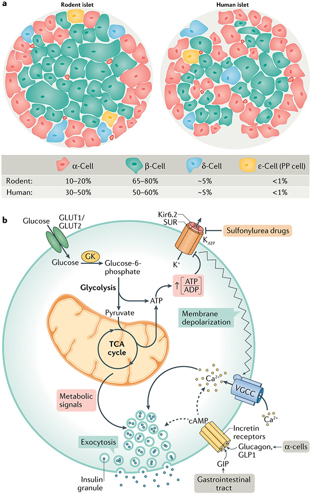
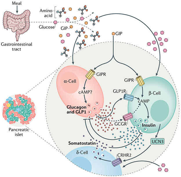
Metabolic homeostasis in mammals is tightly regulated by the complementary actions of insulin and glucagon. The secretion of these hormones from pancreatic β-cells and α-cells, respectively, is controlled by metabolic, endocrine, and paracrine regulatory mechanisms and is essential for the control of blood levels of glucose. The deregulation of these mechanisms leads to various pathologies, most notably type 2 diabetes, which is driven by the combined lesions of impaired insulin action and a loss of the normal insulin secretion response to glucose. Glucose stimulates insulin secretion from β-cells in a bi-modal fashion, and new insights about the underlying mechanisms, particularly relating to the second or amplifying phase of this secretory response, have been recently gained. Other recent work highlights the importance of α-cell-produced proglucagon-derived peptides, incretin hormones from the gastrointestinal tract and other dietary components, including certain amino acids and fatty acids, in priming and potentiation of the β-cell glucose response. These advances provide a new perspective for the understanding of the β-cell failure that triggers type 2 diabetes.


Figures

Fig. 1 ∣
Fig. 1 ∣. Islet cell architecture and fundamental signalling pathways of GSIS.
a ∣ Comparison of the cellular composition of rodent and human islets. Rodent islets are made up of 10–20% of α-cells, found predominantly on the outer mantle, and 65–80% of β-cells comprising their inner core. δ-cells, γ-cells (also known as or pancreatic polypeptide (PP) cells) and ε-cells are found scattered throughout the islet. Human islets contain a higher percentage of α-cells, which are found throughout the islet, and a slightly lower percentage of β-cells than rodents. b ∣ Glucose-stimulated insulin secretion (GSIS) is mediated by a triggering pathway (solid arrows) and amplification pathways (dashed arrows). Glucose uptake into β-cells occurs via GLUT1 (human) or GLUT2 (rodent) transporters; glucose uptake is not rate limiting for GSIS, but rather the rate of β-cell glucose metabolism is controlled by glucokinase (GK), which determines the entry of glucose into the glycolytic pathway, followed by its oxidation via the tricarboxylic acid (TCA) cycle and subsequent generation of ATP. Glucose metabolism initiates the triggering pathway of GSIS via an elevation in the ATP to ADP ratio, resulting in the closure of KATP channels, membrane depolarization and subsequent opening of voltage-gated calcium channels (VGCC). The resulting increase in intracellular calcium drives the triggering phase of insulin granule exocytosis. The importance of GK and the KATP channel for regulating first-phase insulin secretion is clearly illustrated by the functional impact of human genetic mutations in these proteins. Human GK mutations that lower its Km result in a form of persistent hyperinsulinism and hypoglycaemia caused by a lower glucose threshold for the activation of β-cell glucose metabolism and insulin secretion,. Other GK mutations cause partial or complete loss of enzyme activity, resulting in insulin insufficiency and in a form of maturity onset diabetes of the young (MODY2),. Islet cell KATP channels are composed of an octamer of four potassium channel subunits (Kir6.2) and four sulfonylurea receptor 1 (SUR1) regulatory subunits, which bind ATP to cause channel closure, plasma membrane depolarization and subsequent activation of insulin secretion. KATP channel closure can also be induced by sulfonylurea drugs, which are used as anti-diabetic medications. Mutations that force the chronic closure of the channel result in hypoglycaemia due to insulin hypersecretion. Conversely, gain-of-function mutations that prevent closure of the channel by ATP cause syndromes of insulin insufficiency and neonatal diabetes mellitus,. The amplification pathways are dependent upon membrane depolarization initiated by the triggering pathway. Multiple metabolic signalling mechanisms contribute to the amplification of insulin secretion, as detailed in subsequent figures. cAMP, cyclic AMP; GIP, gastric inhibitory polypeptide; GLP1, glucagon-like peptide 1.
Fig. 2 ∣
Fig. 2 ∣. Pyruvate cycling pathways implicated in the regulation of GSIS.
In the post-prandial state, increased anaplerotic metabolism of pyruvate and other fuels leads to the accumulation of tricarboxylic acid (TCA) cycle intermediates, which leave the mitochondria to engage in cytosolic reactions that create signals for the amplifying/second phase of glucose-stimulated insulin secretion (GSIS). This includes (1) the pyruvate–malate cycle (highlighted by the brown circle and involving malic enzyme (ME) and the malate carrier (MC)), (2) the pyruvate–citrate cycle (highlighted by the light blue circle and involving ATP citrate lyase (ACLY), malate dehydrogenase (MDH), the citrate/isocitrate carrier (CIC) and ME), and (3) the pyruvate–isocitrate cycle (highlighted by the green circle and involving the cytosolic NADP-linked isoform of isocitrate dehydrogenase (IDH1), the mitochondrial NADPH-linked isoform of isocitrate dehydrogenase (IDH2) and CIC). Note that the pyruvate–isocitrate pathway is a full cycle that includes reductive TCA flux, involving the carboxylation of α-ketoglutarate (αKG) to isocitrate in the mitochondria by IDH2. In β-cells, this reductive TCA cycle flux ensures that αKG generated by IDH1 is used to regenerate citrate and isocitrate to sustain IDH1 activity. NADPH produced by IDH1 in the cytosol connects to insulin granule exocytosis via glutathione reductase (GSR) to produce reduced glutathione (GSH), leading to the activation of glutaredoxin (GRX1), which mediates the reduction (S2 to SH2 isoform transition) and activation of sentrin/SUMO-specific protease 1 (SENP1). SENP1 then acts as a deSUMOylase that removes SUMO peptides from secretory granule-trafficking proteins to enhance exocytosis. The activity of the pyruvate–isocitrate cycle is maintained by the supply of glutamate, which can be converted to αKG by glutamate dehydrogenase (GDH). The supply of glutamate can be achieved either through the activity of the malate–aspartate shuttle or by the reversible activity of GDH, which is regulated by leucine and its analogue 2-aminobicyclo-(2,2,1)-heptane-2-carboxylic acid (BCH). Recent findings show that the effect of glutamine plus leucine/BCH co-treatment to stimulate insulin secretion is explained, in part, by the isocitrate cycle catalysed by IDH1 and IDH2. Throughout the figure, metabolic enzymes are shown in yellow ovals, whereas mitochondrial organic acid transporters are shown in red ovals. GSSG, glutathione disulfide; OGC, 2-oxoglutarate carrier; PDH, pyruvate dehydrogenase; PC, pyruvate carboxylase.
Fig. 3 ∣
Fig. 3 ∣. Pentose monophosphate shunt, phosphoenolpyruvate cycle and their nucleotide metabolites in GSIS.
As highlighted in the article, the regulation of glucose-stimulated insulin secretion (GSIS) may involve the synthesis of nucleotides other than ATP, specifically adenylosuccinate (S-AMP) generated by the pentose monophosphate shunt or GTP produced in the tricarboxylic acid (TCA) cycle by the GTP-forming isoform of the TCA cycle enzyme succinyl-CoA synthetase (SCS-GTP). Glucose metabolism via the pentose monophosphate shunt generates ribose-5-phosphate and inosine monophosphate (IMP), which is converted to S-AMP via adenylosuccinate synthetase (ADSS). The inhibition of ADSS to block S-AMP formation inhibits GSIS, while infusion of S-AMP into patch-clamped human β-cells amplifies insulin granule exocytosis. NADPH generated in the first two reactions of the pentose monophosphate shunt catalysed by glucose-6-phosphate dehydrogenase (G6PDH) and 6-phosphogluconate dehydrogenase (6PGDH) may synergize with NADPH generated by IDH1 to enhance insulin granule exocytosis via the activation of sentrin/SUMO-specific protease 1 (SENP1) (FIG. 2). The effects of S-AMP on granule exocytosis are attenuated in β-cells with the suppression of SENP1 but the mechanisms by which S-AMP may interact with SENP1 remain unknown (question mark). Alternatively, succinyl-CoA can be converted to succinate in the TCA cycle by SCS-GTP. The manipulation of SCS-GTP expression affects GSIS via the modulation of GTP levels, required for activity of the mitochondrial isoform of phosphoenolpyruvate carboxykinase (PEPCK-M). The synthesis of phosphoenolpyruvate (PEP) by this PEPCK-catalysed PEP cycle produces ATP for the regulation of the KATP channel. OAA, oxaloacetate; PK, pyruvate kinase; PDH, pyruvate dehydrogenase; PC, pyruvate carboxylase.
Fig. 4 ∣
Fig. 4 ∣. Glycerolipid/FFA cycle for the amplification of GSIS.
This model holds that the increase in malonyl-CoA levels induced by stimulatory glucose inhibits carnitine palmitoyl transferase 1 (CPT1) and fatty acid (FA) oxidation, contributing to lipid synthesis and diversion of free fatty acids (FFAs) away from oxidation and towards esterification with glycerol-3-phosphate to form glycerolipids. Glycerolipids are used to form triglycerides, which are then hydrolysed to form diacylglycerols (DAG) and further to monoacylglycerols (MAG), including 1-MAG, which binds the insulin granule trafficking protein Munc13-1 to enhance insulin granule exocytosis. FAs also potentiate glucose-stimulated insulin secretion (GSIS) by interaction with the plasma membrane FFA receptor 1 (FFAR1) to activate the metabolism of phosphoinositide bisphosphate (PIP2) to inositol triphosphate (IP3) and DAG to stimulate insulin secretion via mobilization of Ca2+ and activation of protein kinase C (PKC) and protein kinase D (PKD1), respectively. 1,2-DAG, 1,2-diacylglycerol; ACLY, ATP citrate lyase; FA-CoA, fatty acyl-CoA; LPA, lysophosphatidic acid; OAA, oxaloacetate; PA, phosphatidic acid; TCA, tricarboxylic acid.
Fig. 5 ∣
Fig. 5 ∣. Other hormones in control of insulin secretion.
Nutrient ingestion increases the circulating concentrations of glucose, amino acids and the incretin hormones produced by the gastrointestinal tract: glucose-dependent insulinotropic polypeptide (GIP) and glucagon-like peptide 1 (GLP1). Agonism of either the GIP receptor (GIPR) or GLP1 receptor (GLP1R) potentiates glucose-stimulated insulin secretion (GSIS) through a cyclic AMP (cAMP)-dependent mechanism. GIP also potentiates amino acid-stimulated glucagon secretion in α-cells, which occurs through as-of-yet undefined mechanisms. The secretion of glucagon generally opposes the action of insulin and insulin negatively regulates glucagon secretion. However, there is now evidence that endocrine activity of α-cells is important for regulating glucose-stimulated insulin secretion in β-cells. Together with glucagon, α-cells also produce GLP1 through alternative processesing of the proglucagon peptide precursor, and both hormones increase the level of cAMP in β-cells to regulate insulin secretion in response to a meal. This is mostly mediated by binding of these hormones to GLP1R and to a lesser extent (dashed arrows) to the glucagon receptor (GCGR). Thus, the incretin action of GIP to stimulate insulin secretion includes both direct effects on β-cells and indirect actions on α-cells, mediated by the paracrine stimulatory effects of proglucagon-derived peptides. Urocortin 3 (UCN3) produced by β-cells initiates a separate mechanism of paracrine interaction within the islet. UCN3 enhances δ-cell activity to increase somatostatin secretion through the corticotropin-releasing hormone receptor(CRHR2). The elevation in somatostatin secretion inhibits the secretory activity of β-cells, completing a negative feedback loop.

References

    1. Sakula A Paul Langerhans (1847–1888): a centenary tribute. J. R. Soc. Med 81, 414–415 (1988). - PMC - PubMed
    1. Dolensek J, Rupnik MS & Stozer A Structural similarities and differences between the human and the mouse pancreas. Islets 7, e1024405 (2015). - PMC - PubMed
    1. Ionescu-Tirgoviste C et al. A 3D map of the islet routes throughout the healthy human pancreas. Sci. Rep 5, 14634 (2015). - PMC - PubMed
    1. Olehnik SK, Fowler JL, Avramovich G & Hara M Quantitative analysis of intra- and inter-individual variability of human beta-cell mass. Sci. Rep 7, 16398 (2017). - PMC - PubMed
    1. Saito K, Takahashi T, Yaginuma N & Iwama N Islet morphometry in the diabetic pancreas of man. Tohoku J. Exp. Med 125, 185–197 (1978). - PubMed

Publication types