. 2021 Mar;23(3):187.
doi: 10.3892/mmr.2021.11826. Epub 2021 Jan 5.

Melatonin inhibits proliferation and viability and promotes apoptosis in colorectal cancer cells via upregulation of the microRNA-34a/449a cluster

Affiliations

Melatonin inhibits proliferation and viability and promotes apoptosis in colorectal cancer cells via upregulation of the microRNA-34a/449a cluster

Guangyu Ji et al. Mol Med Rep. 2021 Mar.

Abstract

Colorectal cancer (CRC) has a significant burden on healthcare systems worldwide, and is associated with high morbidity and mortality rates in patients. In 2020, the estimated new cases of colon cancer in the United States are 78,300 in men and 69,650 in women. Thus, developing effective and novel alternative agents and adjuvants with reduced side effects is important to reduce the lethality of the disease and improve the quality of life of patients. Melatonin, a pineal hormone that possesses numerous physiological functions, including anti‑inflammatory and antitumor activities, can be found in various tissues, including the gastrointestinal tract. Melatonin exerts anticarcinogenic effects via various mechanisms; however, the identified underlying molecular mechanisms do not explain the full breadth of anti‑CRC effects mediated by melatonin. MicroRNAs (miRs) serve critical roles in tumorigenesis, however, whether melatonin can inhibit CRC by regulating miRs is not completely understood. In the present study, the roles and mechanism underlying melatonin in CRC were investigated. The proliferation of human CRC cells was tested by CCK8, EDU and colony formation assay. The apoptosis of cancer cells was detected by flow cytometry and western blotting. A xenograft mouse model was constructed and the proliferation and apoptosis of tumor tissue was detected by Ki‑67 and TUNEL staining assay respectively. Reverse transcription‑quantitative PCR and western blotting were performed to measure the regulation of miRs on mRNA, and the dual‑luciferase report analysis experiment was used to verify the direct target genes of miRs. Compared with the control group, melatonin inhibited viability and proliferation, and induced apoptosis in CRC cells. Additionally, the effect of melatonin in a xenograft mouse model was assessed. Compared with the control group, melatonin significantly enhanced the expression levels of the miR‑34a/449a cluster, reduced CRC cell proliferation and viability, and increased CRC cell apoptosis. Finally, the dual‑luciferase reporter assay indicated that Bcl‑2 and Notch1 were the target mRNAs of the miR‑34a/449a cluster. To the best of our knowledge, the present study was the first to suggest that melatonin inhibited proliferation and viability, and promoted apoptosis in CRC cells via upregulating the expression of the miR‑34a/449a cluster in vitro and in vivo. Therefore, melatonin may serve as a potential therapeutic for CRC.

Keywords: melatonin; colorectal cancer; microRNA‑34a/449a cluster; proliferation; apoptosis.


Figures
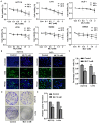
Figure 1.
Figure 1.
Melatonin suppresses CRC cell viability and proliferation. (A) HCT116, LoVo, SW480, SW620, HT-29 and DLD-1 CRC cells were treated with 0.1, 0.5, 1, 1.5 or 2 mM melatonin for 24 or 48 h. Cell viability was assessed by performing an MTT assay. *P<0.05 vs. 24 h control; #P<0.05 vs. 48 h control. Following treatment with melatonin for 48 h, HCT116 and LoVo CRC cell proliferation was assessed by (B) performing EdU staining (scale bar, 50 µm) and (C) quantified by counting the number of EdU+ cells. Following treatment with melatonin for 48 h, CRC cell proliferation was assessed by (D) performing colony formation assays and (E) quantified. *P<0.05 vs. control. Data are presented as the mean ± standard deviation. CRC, colorectal cancer; Con, control; MLT, melatonin.
Figure 2.
Figure 2.
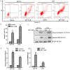
Melatonin induces CRC cell apoptosis. HCT116 and LoVo CRC cells were treated with 2 mM melatonin for 48 h. Cell apoptosis was (A) determined via flow cytometry following Annexin V/PI staining and (B) quantified. Cleaved caspase-3 and cleaved PARP protein expression levels were (C) determined via western blotting and (D) semi-quantified. *P<0.05 vs. control. Data are presented as the mean ± standard deviation. CRC, colorectal cancer; PARP, poly(ADP-ribose) polymerase 1; MLT, melatonin; Con, control.
Figure 3.
Figure 3.
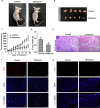
Melatonin inhibits tumor growth in vivo. Representative images of (A) tumor-bearing mice and (B) tumors on the day of harvesting. Tumor (C) volume and (D) weight. (E) Representative images of HE staining (scale bar, 50 µm). (F) Ki-67 staining was conducted to assess tumor cell proliferation (scale bar, 100 µm). (G) TUNEL staining was performed to assess tumor cell apoptosis (scale bar, 50 µm). *P<0.05 vs. control. Data are presented as the mean ± standard deviation. HE, hematoxylin and eosin.
Figure 4.
Figure 4.
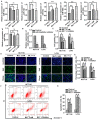
Melatonin affects CRC cell proliferation and apoptosis by regulating the expression of the miR-34a/449a cluster. HCT116 cells were treated with 2 mM melatonin for 48 h. (A) The expression levels of miR-125a, miR-365a, miR-34a, miR-449a, miR-184 and miR-143a was determined via reverse transcription-quantitative PCR. (B) The transfection efficiency of miR-34a/449a inhibitor in HCT116 and LoVo cells. *P<0.05 vs. control. (C) HCT116 and LoVo cell viability were detected using a Cell Counting Kit-8 assay. CRC cell proliferation was (D) determined by performing EdU staining (scale bar, 50 µm) and (E) the number of EdU+ cells was quantified. CRC cell apoptosis was (F) determined via flow cytometry and (G) quantified. *P<0.05, **P<0.01 and #P<0.05 vs. MLT 2 mM. Data are presented as the mean ± standard deviation. CRC, colorectal cancer; miR, microRNA; ns, not significant; MLT, melatonin.
Figure 5.
Figure 5.
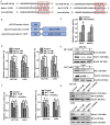
Bcl-2 and Notch1 are direct target genes of the miR-34a/449a cluster. (A) The predicted binding sequences between miR-34a/449a cluster and the 3′UTRs of Bcl-2 and Notch1. (B) Schematic illustration of the pGL3-promoter luciferase reporter constructs that were used for examining the effect of the miR-34a/449a cluster on the 3′UTRs of Bcl-2 and Notch1. (C) The dual luciferase reporter assay was performed to verify the interaction between the miR-34a/449a cluster and the 3′UTRs of Bcl-2 and Notch1. HCT116 and LoVo cells were treated with 1 or 2 mM melatonin for 48 h. Bcl-2 and Notch1 (D) mRNA and (E) protein expression levels were determined via RT-qPCR and western blotting, respectively. HCT116 and LoVo cells were pretreated with the miR-34a-449a cluster inhibitor and then treated with 2 mM melatonin for 48 h. Bcl-2 and Notch1 (F) mRNA and (G) protein expression levels were determined via RT-qPCR and western blotting, respectively. *P<0.05 vs. control; #P<0.05 vs. 2 mM MLT. Data are presented as the mean ± standard deviation. miR, microRNA; UTR, untranslated region; RT-qPCR, reverse transcription-quantitative PCR; MLT, melatonin; Luc, luciferase.
Figure 6.
Figure 6.
Schematic diagram of the mechanism underlying melatonin-mediated inhibition of proliferation and induction of apoptosis in colorectal cancer. miRNA, microRNA.

References

    1. Chen W, Zheng R, Baade PD, Zhang S, Zeng H, Bray F, Jemal A, Yu XQ, He J. Cancer statistics in China, 2015. CA Cancer J Clin. 2016;66:115–132. doi: 10.3322/caac.21338. - DOI - PubMed
    1. Siegel RLM, Miller KD, Jemal A. Cancer statistics, 2020. CA Cancer J Clin. 2020;70:7–30. doi: 10.3322/caac.21601. - DOI - PubMed
    1. Zhang Y, Chen Z, Li J. The current status of treatment for colorectal cancer in China: A systematic review. Medicine (Baltimore) 2017;96:e8242. doi: 10.1097/MD.0000000000008242. - DOI - PMC - PubMed
    1. Scholefield JH, Steele RJ, British Society For Gastroenterology; Association of Coloproctology for Great Britain and Ireland Guidelines for follow up after resection of colorectal cancer. Gut. 2002;51(Suppl 5):V3–V5. doi: 10.1136/gut.51.suppl_5.v3. - DOI - PMC - PubMed
    1. Cunningham D, Humblet Y, Siena S, Khayat D, Bleiberg H, Santoro A, Bets D, Mueser M, Harstrick A, Verslype C, et al. Cetuximab monotherapy and cetuximab plus irinotecan in irinotecan-refractory metastatic colorectal cancer. N Engl J Med. 2004;351:337–345. doi: 10.1056/NEJMoa033025. - DOI - PubMed