. 2021 Apr;155(4):477-489.
doi: 10.1007/s00418-020-01949-8. Epub 2021 Jan 4.

A combinatorial method to visualize the neuronal network in the mouse spinal cord: combination of a modified Golgi-Cox method and synchrotron radiation micro-computed tomography

Affiliations

A combinatorial method to visualize the neuronal network in the mouse spinal cord: combination of a modified Golgi-Cox method and synchrotron radiation micro-computed tomography

Liyuan Jiang et al. Histochem Cell Biol. 2021 Apr.

Abstract

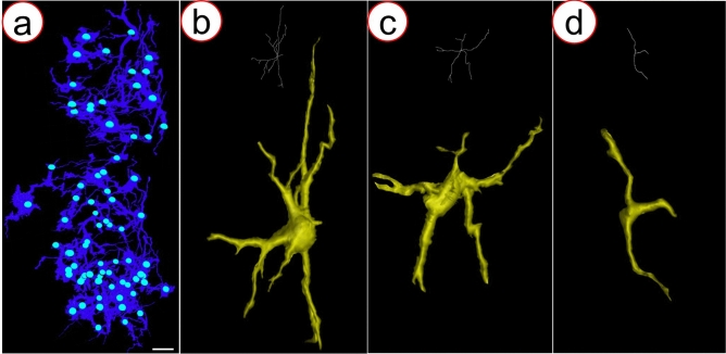
Exploring the three-dimensional (3D) morphology of neurons is essential to understanding spinal cord function and associated diseases comprehensively. However, 3D imaging of the neuronal network in the broad region of the spinal cord at cellular resolution remains a challenge in the field of neuroscience. In this study, to obtain high-resolution 3D imaging of a detailed neuronal network in the mass of the spinal cord, the combination of synchrotron radiation micro-computed tomography (SRμCT) and the Golgi-cox staining were used. We optimized the Golgi-Cox method (GCM) and developed a modified GCM (M-GCM), which improved background staining, reduced the number of artefacts, and diminished the impact of incomplete vasculature compared to the current GCM. Moreover, we achieved high-resolution 3D imaging of the detailed neuronal network in the spinal cord through the combination of SRμCT and M-GCM. Our results showed that the M-GCM increased the contrast between the neuronal structure and its surrounding extracellular matrix. Compared to the GCM, the M-GCM also diminished the impact of the artefacts and incomplete vasculature on the 3D image. Additionally, the 3D neuronal architecture was successfully quantified using a combination of SRμCT and M-GCM. The SRμCT was shown to be a valuable non-destructive tool for 3D visualization of the neuronal network in the broad 3D region of the spinal cord. Such a combinatorial method will, therefore, transform the presentation of Golgi staining from 2 to 3D, providing significant improvements in the 3D rendering of the neuronal network.

Keywords: Modified Golgi-Cox method; Neuronal network; SRμCT; Spinal cord; Three-dimension.


Conflict of interest statement

The authors declare no conflict of interest.

Figures

Fig. 1
Fig. 1
Schematic depiction of beamline experimental station at the BL13W1 at the Shanghai Synchrotron Radiation Facility (SSRF) in China. The samples were fixed on a sample rotation stage. The images were collected by a detector located at a 3.5 cm distance from the sample after transmission of a monochromatic synchrotron radiation X-ray beam through the sample and delivery to the image acquisition system
Fig. 2
Fig. 2
Comparison of photomicrographs from the GCM group and M-GCM group. ac Representative images of the fresh and stained spinal cord tissue M-GCM group (a, b, and c are 4×, 10×, and 20× image, respectively). df Representative images of the fresh and stained spinal cord tissue GCM group (d, e, and f are 4×, 10×, and 20× image, respectively). Red arrows indicate the artefacts and the green arrows indicate the incomplete vascular structure. g Representative 60 × image shows low background staining in the M-GCM group. h Representative 60 × image shows high background staining in the GCM group. i A histogram illustrating M-GCM has significantly less the gray value of background compared to that of GCM. j Reconstructive 3D images of neuronal networks in the M-GCM group. k Reconstructive 3D images of neuronal networks in the GCM group. l A histogram illustrating M-GCM has significantly artefact in tissue compared to that of GCM. ***P < 0.0001, Student’s t test was used to determine the statistical significance of the differences [scale bar: 120 μm (g, h) and 250 μm (j, k)]
Fig. 3
Fig. 3
Projection images detected by a high-resolution SRμCT. a A representative projection image of a stained spinal cord from the M-GCM group. b Local magnification of the region of interest is denoted by the red frame in a. d A representative projection images of a spinal cord from the GCM group. e Local magnification of the region of interest is denoted by the red frame in d. c Profile of the grey value along the red line in b and e. f A histogram illustrating M-GCM achieved a significantly higher contrast between the neuronal site and surrounding background compared to GCM did. ***P < 0.0001, Student’s t test was used to determine the statistical significance of the differences (scale bar: 200 μm)
Fig. 4
Fig. 4
3D images of the neuronal network by SRμCT. a, c A representative 3D image of the neuronal network from the M-GCM group. b, d A corresponding 3D image of the neuronal network from the GCM group. The green arrows indicate the incomplete vascular structure [scale bar: 250 μm (a, b) and 100 μm (c, d)]
Fig. 5
Fig. 5
Images of the neuronal network in the ventral horn of the mouse spinal cord. a Photomicrograph of the neuronal network in the ventral horn. b Digital 3D image of the neuronal network in the ventral horn from the M-GCM group. c Digital 3D image of the neuronal network in the ventral horn from the GCM group (scale bar: 50 μm)
Fig. 6
Fig. 6
Images of motor neurons in the mouse spinal cord. a Schematic depiction of a neuron. b Photomicrograph of the motor neurons. c 3D reconstructive motor neurons from the M-GCM group. d 3D reconstructive motor neurons from the GCM group. Red arrows indicate the axons, green arrows indicate dendrites, and yellow arrows indicate soma. A Histogram illustrating M-GCM visualized longed neurite than GCM did. ***P < 0.0001, Student’s t test was used to determine the statistical significance of the differences (scale bar: 50 μm)
Fig. 7
Fig. 7
a 3D neuronal network at the thoracic mouse spinal cord, quantification for soma number. bd Neurons with different shapes, measurements of neurites length. As the figure is shown, the combination of SRμCT and M-GCM has an advantage in the quantification of detailed neuronal architecture (scale bar: 50 μm)
Fig. 8
Fig. 8
a The representative image of stained neurons in the unscanned spinal cord tissue; b the representative image of stained neurons in the scanned spinal cord tissue (scale bar: 100 μm)

Similar articles

Cited by

References

    1. Bentivoglio M, Cotrufo T, Ferrari S, Tesoriero C, Mariotto S, Bertini G, Berzero A, Mazzarello P. The original histological slides of camillo golgi and his discoveries on neuronal structure. Front Neuroanat. 2019;13:3. doi: 10.3389/fnana.2019.00003. - DOI - PMC - PubMed
    1. Bukreeva I, Campi G, Fratini M, Spano R, Bucci D, Battaglia G, Giove F, Bravin A, Uccelli A, Venturi C, Mastrogiacomo M, Cedola A. Quantitative 3D investigation of Neuronal network in mouse spinal cord model. Sci Rep. 2017;7:41054. doi: 10.1038/srep41054. - DOI - PMC - PubMed
    1. Cao Y, Wu T, Yuan Z, Li D, Ni S, Hu J, Lu H. Three-dimensional imaging of microvasculature in the rat spinal cord following injury. Sci Rep. 2015;5:12643. doi: 10.1038/srep12643. - DOI - PMC - PubMed
    1. Cao Y, Zhou Y, Ni S, Wu T, Li P, Liao S, Hu J, Lu H. Three dimensional quantification of microarchitecture and vessel regeneration by synchrotron radiation microcomputed tomography in a rat model of spinal cord injury. J Neurotrauma. 2017;34(6):1187–1199. doi: 10.1089/neu.2016.4697. - DOI - PubMed
    1. Cedola A, Bravin A, Bukreeva I, Fratini M, Pacureanu A, Mittone A, Massimi L, Cloetens P, Coan P, Campi G, Spano R, Brun F, Grigoryev V, Petrosino V, Venturi C, Mastrogiacomo M, Kerlero de Rosbo N, Uccelli A. X-ray phase contrast tomography reveals early vascular alterations and neuronal loss in a multiple sclerosis model. Sci Rep. 2017;7(1):5890. doi: 10.1038/s41598-017-06251-7. - DOI - PMC - PubMed

LinkOut - more resources