. 2021 Jan 20;143(2):891-901.
doi: 10.1021/jacs.0c10696. Epub 2021 Jan 5.

Targeting the APP-Mint2 Protein-Protein Interaction with a Peptide-Based Inhibitor Reduces Amyloid-β Formation

Affiliations

Targeting the APP-Mint2 Protein-Protein Interaction with a Peptide-Based Inhibitor Reduces Amyloid-β Formation

Christian R O Bartling et al. J Am Chem Soc. .

Abstract

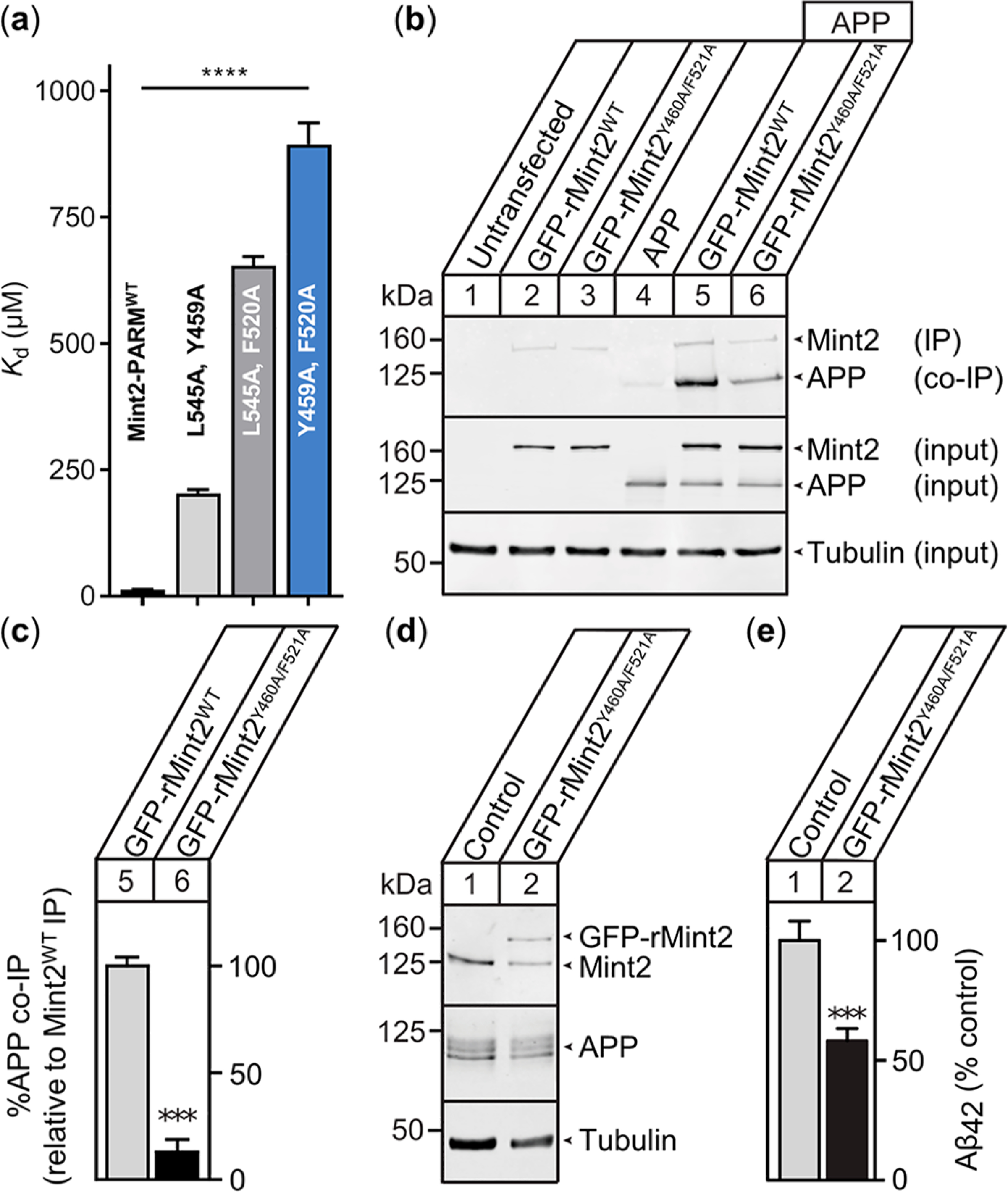
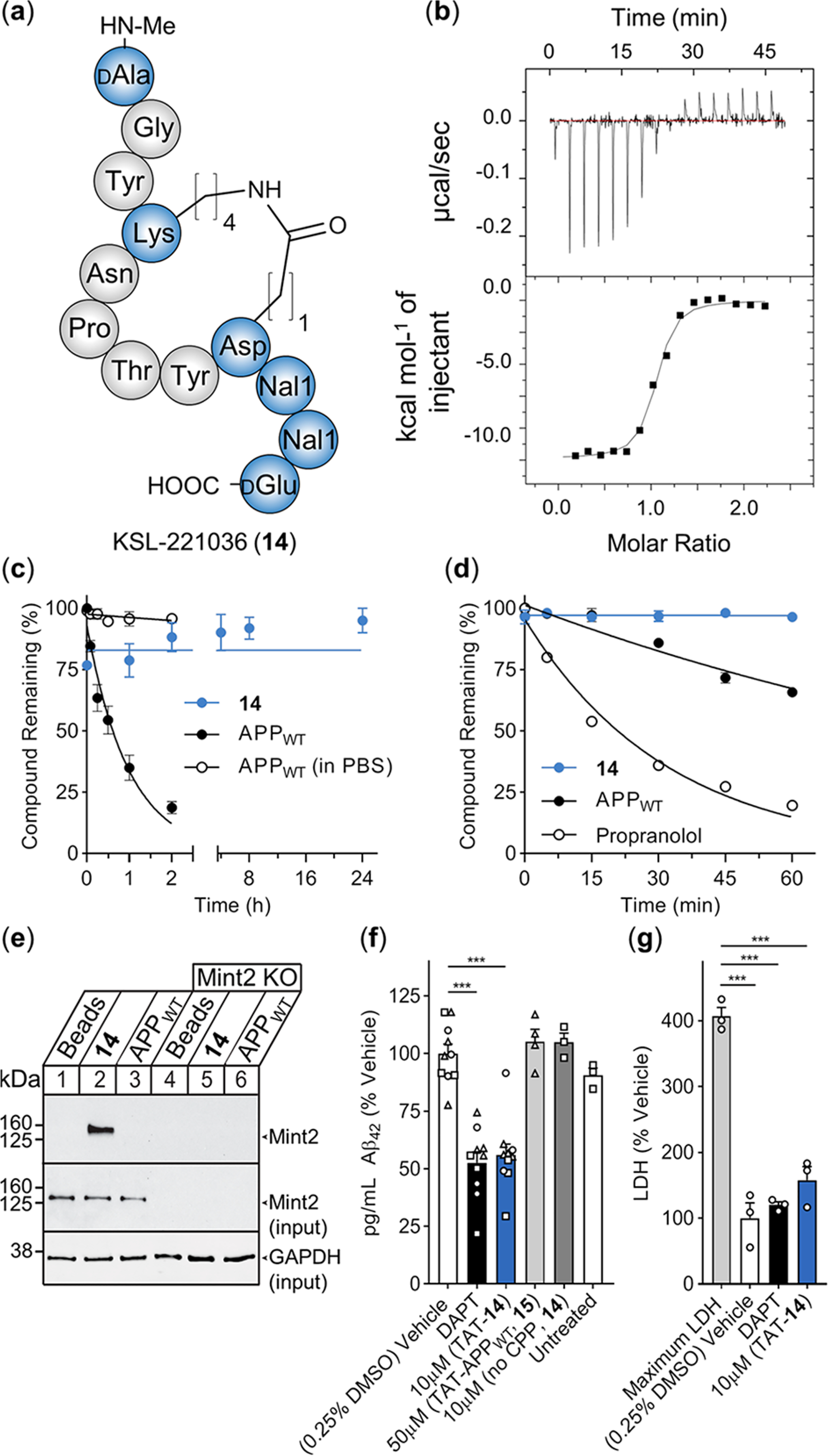
There is an urgent need for novel therapeutic approaches to treat Alzheimer's disease (AD) with the ability to both alleviate the clinical symptoms and halt the progression of the disease. AD is characterized by the accumulation of amyloid-β (Aβ) peptides which are generated through the sequential proteolytic cleavage of the amyloid precursor protein (APP). Previous studies reported that Mint2, a neuronal adaptor protein binding both APP and the γ-secretase complex, affects APP processing and formation of pathogenic Aβ. However, there have been contradicting results concerning whether Mint2 has a facilitative or suppressive effect on Aβ generation. Herein, we deciphered the APP-Mint2 protein-protein interaction (PPI) via extensive probing of both backbone H-bond and side-chain interactions. We also developed a proteolytically stable, high-affinity peptide targeting the APP-Mint2 interaction. We found that both an APP binding-deficient Mint2 variant and a cell-permeable PPI inhibitor significantly reduced Aβ42 levels in a neuronal in vitro model of AD. Together, these findings demonstrate a facilitative role of Mint2 in Aβ formation, and the combination of genetic and pharmacological approaches suggests that targeting Mint2 is a promising therapeutic strategy to reduce pathogenic Aβ levels.


Conflict of interest statement

The authors declare no competing financial interest.

Figures

Figure 1.
Figure 1.
Structural overview of the Mint protein family and the Mint2-APP interaction. (a) Schematic architecture of Mint proteins illustrating their conserved C-terminal region consisting of a phosphotyrosine binding (PTB) domain (gray), an α-helical ARM linker (dark gray), two PSD-95/drosophila discs large/zonula occludens (ZO-1) (PDZ) domains (light gray), and a variable N-terminal region. Numbering corresponds to human residues. MI = Munc-18 interaction domain, CI = CASK interaction domain. (b) Cartoon representation of the interaction between rat Mint2-PARM (gray) and APP (residues 754–767; blue). The APPC-term is binding at the interface between the α3-helix and β5-strand of Mint2, forming an antiparallel β-sheet (N-terminal) followed by a β-turn and a C-terminal α-helix. The ARM linker (dark gray) is found in an open conformation enabling APP binding. (c) Structure of the APP peptide (blue sticks) bound to the rat Mint2-PTB domain (gray). The β5-strand of the PTB domain (gray stick, side chains not depicted) is shown to highlight the backbone–backbone H-bond network (black dotted lines) between the Mint2-PTB domain and APP (PDB ID: 3SV1).
Figure 2.
Figure 2.
Semisynthesis of Mint2-PARM and effects of A-to-E and Ala mutations in APP and Mint2-PARM. (a) Semisynthetic approach to introducing A-to-E substitutions in Mint2-PARM. N-terminal fragment PARMN (A479-Q570) is fused to a Xa site to generate an N-terminal Cys by factor Xa cleavage. C-terminal fragment PARMC (E364-H452) is expressed with a C-terminal intein to generate the required C-terminal thioester. Semisynthesis is initiated by ligating synthetic peptide fragment PARMpep to PARMN. Next, the Thz group is converted to a free Cys and PARMpep+N is ligated to PARMC. Semisynthetic Mint2-pPARMSS is obtained after desulfurization and refolding (Figures S2–S7). (b) Affinity fold-change of the APPWT peptide’s Ala-scan and (c) A-to-E substitutions toward Mint2-PARM obtained in an inhibition FP assay (Ki) reported relative to the APPWT peptide [Ki(mutant)/Ki(APPWT) + SEM]. (d) Affinity fold change of the APPC‑term peptide binding to Mint2-PARM Ala and (e) A-to-E variants obtained by the saturation FP assay (Kd) relative to Mint2-PARMWT [Kd(variant)/Kd(wild type) + SEM]. See Figures S8 and S9 for FP data.
Figure 3.
Figure 3.
APP binding-deficient rMint2Y460A/F521A variant reduces Aβ42 levels in primary mouse neurons. (a) APPC‑term peptide affinity for selected Mint2-PARM variants. Data are expressed as the mean + SEM (n = 3). The statistical significance was evaluated using one-way ANOVA with Dunnett’s multiple comparison test, **** P < 0.0001 (Figure S10). (b) Coimmunoprecipitation of APP with the Mint2 antibody from HEK293T cells cotransfected with APP and GFP-rMint2WT or GFP-rMint2Y460A/F521A. Western blotting indicates less APP binding to rMint2Y460A/F521A (lane 6) than to GFP-Mint2WT (lane 5). α-Tubulin served as a loading control. (c) Quantification of APP coimmunoprecipitation using GFP-rMint2WT and GFP-rMint2Y460A/F521A. Data were normalized to GFP-rMint2WT and expressed as the mean + SEM. The statistical significance was evaluated with the Student’s t-test, *** P < 0.001. (n = 4 independent experiments). (d) Western blot for Mint2, APP, and α-tubulin from neuronal lysates carrying the APPswe/PS1ΔE9 mutation and infected with the lentiviral GFP-rMint2Y460A/F521A mutant. (e) Aβ42 ELISA quantification of conditioned media from neurons overproducing Aβ shows reduced Aβ42 levels when neurons were infected with the GFP-rMint2Y460A/F521A mutant. Data were normalized to the endogenous Mint2WT control and expressed as the mean + SEM (n = 7 biological replicates from two independent experiments). The statistical significance was evaluated using the Student’s t-test, *** P < 0.001.
Figure 4.
Figure 4.
Incorporation of ncAAs and evaluation of side chain-to-side chain macrocyclization in the APPWT peptide. (a) Overview of synthesized APPWT peptide variants, including the structure of the introduced amino acids for each position (blue). (b) Ki values of each APPWT peptide variant measured by FP. n.b. indicates nonbinding peptide (i.e., Ki ≥ 500 μM). Data are expressed as the mean + SEM (n = 3). (c) Structure of side chain-to-side chain cyclized APPWT peptide analogues with native residue order (Glu in the i position, dark gray) and (d) inverse residue order (Lys in the i position, light gray). (e) Ki values of cyclic APPWT peptide variants measured by FP. Data are expressed as the mean + SEM (n = 3). (f) Helical propensity of residues Y762 to E766 from the molecular dynamics simulation of cyclic APPWT peptide variants 1 and 3–13 (% helical frames) plotted against the binding affinity (the mean Ki value) including the trend line (blue).
Figure 5.
Figure 5.
PPI inhibitor of the APP-Mint2 interaction reduces Aβ42 production in the neuronal in vitro model of AD. (a) Schematic structure of KSL-221036 (14). (b) Representative ITC of titrating Mint2-PARM with 14; raw heat signature (top) and integrated molar heat release (bottom). (c) In vitro plasma stability of 14 (blue) and the APPWT peptide (black) including buffer control for the APPWT peptide (circles); data are expressed as the mean ± SEM (n = 3). (d) In vitro hepatic clearance of 14 (blue) and the APPWT peptide (black) including propranolol control (circles, n = 1); data are expressed as the mean ± SEM (n = 3). (e) Representative pull down of Mint2 from neuronal cell lysate (15 DIV). Lanes 1–3: expression of Mint2. Lanes 4–6: knockout of Mint2. The eluent was resolved by SDS-PAGE and immunoblotted for Mint2 and GAPDH (n = 3). (f) Aβ42 ELISA quantification from conditioned media of neurons overproducing Aβ; data are normalized to the vehicle control and expressed as the mean + SEM (n = 3 independent experiments, n = 10 biological replicates for the vehicle, DAPT, and TAT-14 (10 μM). Independent experiments are represented by different shapes. Statistical significance evaluated using one-way ANOVA with Dunnett’s multiple comparison test, ***P < 0.001. (g) LDH levels from conditioned medium of cultured neurons overproducing Aβ collected after 24 h of treatment with DAPT and TAT-14 (10 μM). Maximum LDH represents the maximum amount of LDH released from neurons lysed using detergent. Individual data points in (g) correspond to data in panel (f) (circles = same assay). Data are expressed as %LDH normalized to vehicle control and shown as the mean + SEM. Statistical significance was evaluated using one-way ANOVA with Sidak’s multiple comparison test, ***P < 0.001.

References

    1. Wang J; Gu BJ; Masters CL; Wang YJ A systemic view of Alzheimer disease - insights from amyloid-beta metabolism beyond the brain. Nat. Rev. Neurol. 2017, 13 (10), 612–623. - PubMed
    1. Selkoe DJ; Hardy J The amyloid hypothesis of Alzheimer’s disease at 25 years. EMBO Mol. Med. 2016, 8 (6), 595–608. - PMC - PubMed
    1. Müller UC; Deller T; Korte M Not just amyloid: physiological functions of the amyloid precursor protein family. Nat. Rev. Neurosci. 2017, 18 (5), 281–298. - PubMed
    1. Haass C; Selkoe DJ Soluble protein oligomers in neurodegeneration: lessons from the Alzheimer’s amyloid beta-peptide. Nat. Rev. Mol. Cell Biol. 2007, 8 (2), 101–112. - PubMed
    1. Panza F; Lozupone M; Logroscino G; Imbimbo BP A critical appraisal of amyloid-beta-targeting therapies for Alzheimer disease. Nat. Rev. Neurol. 2019, 15 (2), 73–88. - PubMed

Publication types

MeSH terms

LinkOut - more resources