. 2020 Oct;9(10):731-741.
doi: 10.1302/2046-3758.910.BJR-2019-0368.R2.

Less mechanical loading attenuates osteoarthritis by reducing cartilage degeneration, subchondral bone remodelling, secondary inflammation, and activation of NLRP3 inflammasome

Affiliations

Less mechanical loading attenuates osteoarthritis by reducing cartilage degeneration, subchondral bone remodelling, secondary inflammation, and activation of NLRP3 inflammasome

Zhennian He et al. Bone Joint Res. 2020 Oct.

Abstract

Aims: Osteoarthritis (OA) is a disabling joint disorder and mechanical loading is an important pathogenesis. This study aims to investigate the benefits of less mechanical loading created by intermittent tail suspension for knee OA.

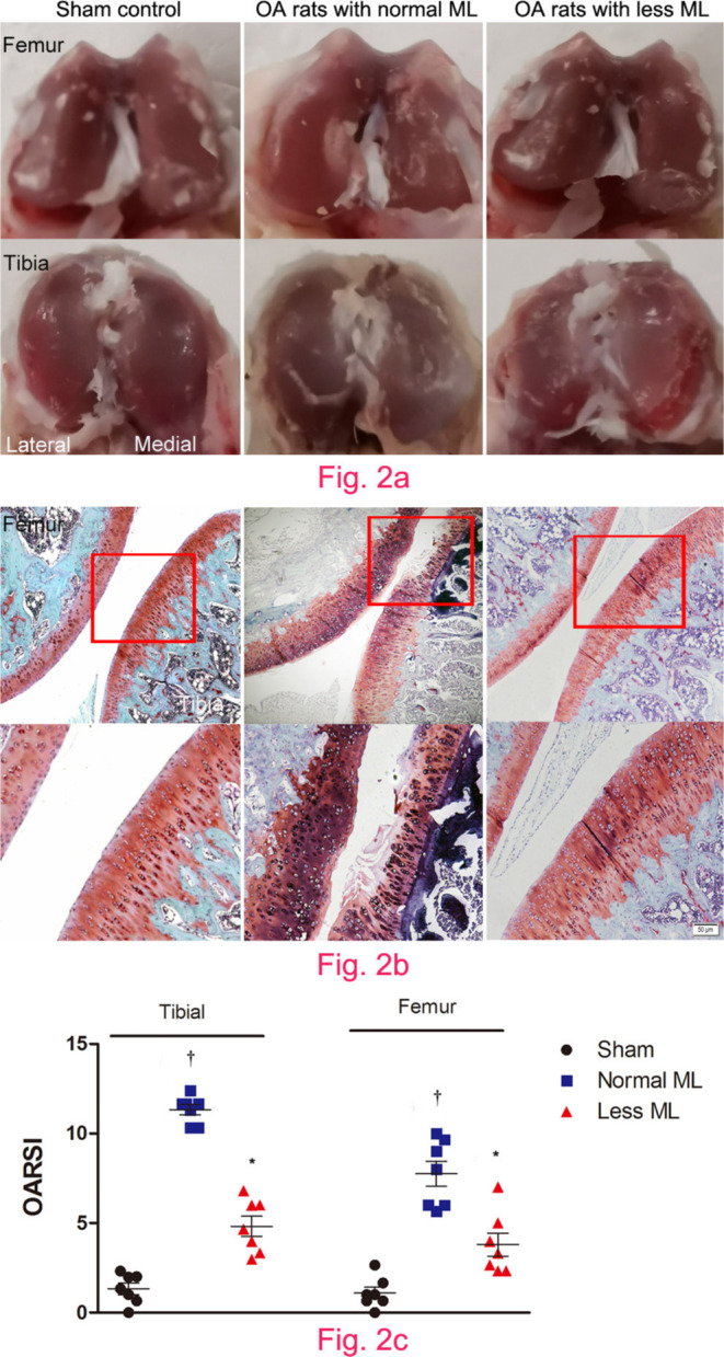
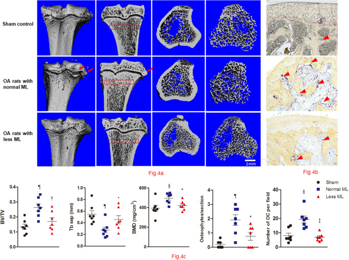
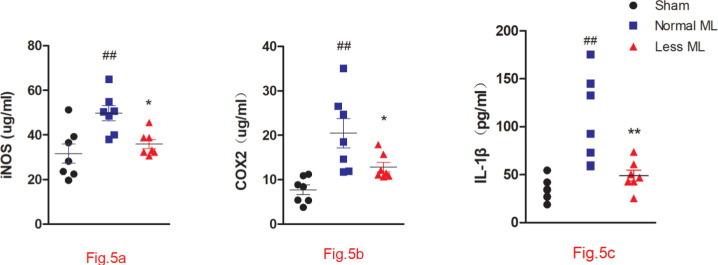
Methods: A post-traumatic OA model was established in 20 rats (12 weeks old, male). Ten rats were treated with less mechanical loading through intermittent tail suspension, while another ten rats were treated with normal mechanical loading. Cartilage damage was determined by gross appearance, Safranin O/Fast Green staining, and immunohistochemistry examinations. Subchondral bone changes were analyzed by micro-CT and tartrate-resistant acid phosphatase (TRAP) staining, and serum inflammatory cytokines were evaluated by enzyme-linked immunosorbent assay (ELISA).

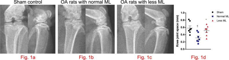
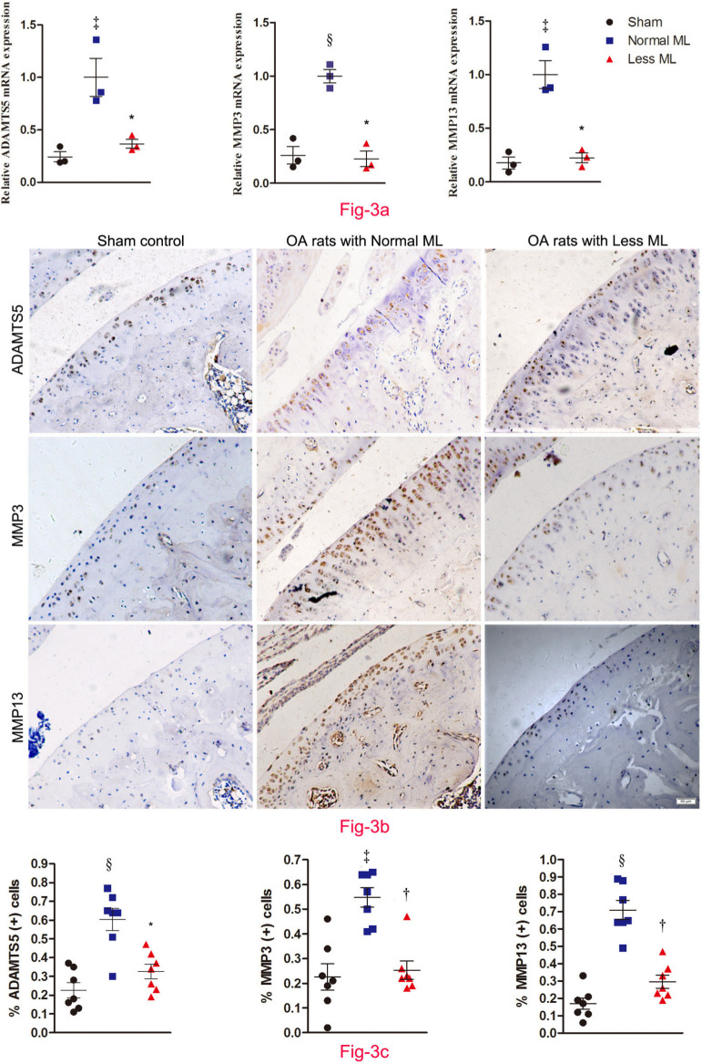
Results: Our radiographs showed that joint space was significantly enlarged in rats with less mechanical loading. Moreover, cartilage destruction was attenuated in the less mechanical loading group with lower histological damage scores, and lower expression of a disintegrin and metalloproteinase with thrombospondin motifs (ADAMTS)-5, matrix metalloproteinase (MMP)-3, and MMP-13. In addition, subchondral bone abnormal changes were ameliorated in OA rats with less mechanical loading, as reduced bone mineral density (BMD), bone volume/tissue volume (BV/TV), and number of osteophytes and osteoclasts in the subchondral bone were observed. Finally, the level of serum inflammatory cytokines was significantly downregulated in the less mechanical loading group compared with the normal mechanical loading group, as well as the expression of NACHT, LRR, and PYD domains-containing protein 3 (NLRP3), caspase-1, and interleukin 1β (IL-1β) in the cartilage.

Conclusion: Less mechanical loading alleviates cartilage destruction, subchondral bone changes, and secondary inflammation in OA joints. This study provides fundamental insights into the benefit of non-weight loading rest for patients with OA. Cite this article: Bone Joint Res 2020;9(10):731-741.

Keywords: Cartilage destruction; Mechanical loading; NLRP3 inflammasome; Osteoarthritis; Subchondral bone.


Figures

Fig. 1
Fig. 1
Less mechanical loading (ML) strategy enlarging the knee joint space in an osteoarthritis (OA) rat. a) The frontal and lateral radiographs of knee joint in sham control. As compared with the narrowed and indistinct joint space in OA rats with normal ML (arrow, b), the joint space was enlarged in OA rats with less ML (arrow, c). d) Furthermore, joint space, as measured in the frontal images, enlarged significantly in rats with less ML. One-way analysis of variance (ANOVA) was used, *p < 0.05 between OA rats with normal and less ML. †p < 0.01 between sham controls and OA rats with normal ML.
Fig. 2
Fig. 2
Less mechanical loading (ML) attenuated articular cartilage degeneration in an osteoarthritis (OA) rat. a) Gross appearance of the articular cartilage in the normal ML group showed that joint surface was rough with focal defects in the proximal medial tibial plateau (lower panel), while smooth joint surface was present in the less ML joint articular cartilage. b) Safranin O and Fast Green staining demonstrated that there were some clefts and breakdown in the cartilage in OA rats with normal ML. a) and b) However, articular surface was relatively smooth and there was no evidence of breakdown in OA rats in the less ML group. c) Furthermore, the Osteoarthritis Research Society International (OARSI) scores in both tibial and femur cartilage were greater in the OA rats with normal ML than that of OA rats with less ML. Scale bar: 50 μm. Values are presented as mean and standard error of the mean (SEM). One-way analysis of variance (ANOVA) was used, *p < 0.001 between OA rats with normal and less ML. †p < 0.001 between sham controls and OA rats with normal ML. Magnification of upper panel in Fig. 2b was 100×, and lower panel was 400×.
Fig. 3
Fig. 3
Less mechanical loading (ML) ameliorates joint catabolism in an osteoarthritis (OA) rat. a) Quantitative polymerase chain reaction (qPCR) revealed that the messenger RNA (mRNA) expression of a disintegrin and metalloproteinase with thrombospondin motifs (ADAMTS)-5, matrix metalloproteinase (MMP)-3, and MMP-13 in the less ML group was lower than that of the normal ML group. b) Furthermore, immunohistochemical examination showed that there were numerous ADAMTS-5, MMP-3, and MMP-13 positive chondrocytes in both the superficial layer and deep layer of cartilage from OA rats with normal ML, while they were seldom expressed in the superficial cartilage in the less ML group. The presentative images were selected from weight-bearing area of tibial cartilage. c) In addition, quantitative analysis demonstrated that the percentage of target positive chondrocytes in articular cartilage was significantly reduced in the less ML group, compared to the normal ML group. Scale bar: 50 μm. Values are presented as mean and standard error of the mean (SEM). One-way analysis of variance (ANOVA) was used, *p < 0.01; †p < 0.001, between OA rats with normal and less ML. ‡p < 0.01; §p < 0.001, between sham controls and OA rats with normal ML. Magnification: 200×.
Fig. 4
Fig. 4
Less mechanical loading (ML) reduced abnormal bone remodelling in subchondral bone in an osteoarthritis (OA) rat. a) Micro-CT of the tibia subchondral bone of rats showed that OA rats with less ML displayed decreased bone mineral density (BMD) and increased trabecular separation (TbSp), compared with OA rats with normal ML. b) The osteophytes were counted in three sections of each rat, and findings revealed that the mean number of osteophytes per joint in the less ML group was significantly lower than that of the normal ML group (red arrows). In addition, OA rats with less ML also showed a significantly lower number of osteoclasts (OCs) per bone volume, compared with the normal ML group (red arrowheads). c) Furthermore, quantitative analysis showed that OA rats with less ML had significantly lower bone volume/tissue volume (BV/TV) and BMD, and greater TbSep, which was seen in the subchondral bone 200 μm to 400 μm below the growth plate (red boxes in a). Scale bar: 1 mm. Values are presented as mean and standard error of the mean (SEM). One-way analysis of variance (ANOVA) was used, *p < 0.05; †p < 0.01; ‡p < 0.001, between OA rats with normal and less ML. §p < 0.01; ¶p < 0.001, between sham controls and OA rats with normal ML. Magnification of Fig. 4b: 200×.
Fig. 5
Fig. 5
Less mechanical loading (ML) ameliorates joint inflammation in an osteoarthritis (OA) rat. Enzyme-linked immunosorbent assay (ELISA) experiment revealed that the concentrations of a) cyclooxygenase-2 (COX-2), b) inducible nitric oxide synthase (iNOS), and c) interleukin 1β (IL-1β) in the less ML group were significantly lower than those in the normal ML group. Values are presented as mean and standard error of the mean (SEM). One-way analysis of variance (ANOVA) was used, *p < 0.05; †p < 0.01, between OA rats with normal and less ML; ‡p < 0.01, between sham controls and OA rats with normal ML.
Fig. 6
Fig. 6
Less mechanical loading (ML) reduced the NACHT, LRR, and PYD domains-containing protein 3 (NLRP3)/caspase 1/interleukin 1β (IL-1β) expression in the cartilage of an osteoarthritis (OA) rat. a) The transcriptional level of NLRP3, caspase-1, and IL-1β was lower in the less ML group than in the normal ML group. b) Furthermore, extensive immunostaining of NLRP3, caspase-1, and IL-1β in normal ML OA cartilage was noted, whereas fewer immune-positive cells could be found in the less ML OA cartilage. The presentative images were selected from weight-bearing area of tibial cartilage. c) The percentage of immunopositive cells of NLRP3, caspase-1, and IL-1β in the less ML group was significantly lower than that of the normal ML group. Scale bar: 50 μm. Values are presented as mean standard error of the mean (SEM). One-way analysis of variance (ANOVA) was used, *p < 0.05; †p < 0.01; ‡p < 0.001, between OA rats with normal and less ML. §p < 0.05; ¶p < 0.01; **p < 0.001, between sham controls and OA rats with normal ML. Magnification in Fig. 6b: 200×.

References

    1. Martel-Pelletier J, Boileau C, Pelletier J-P, Roughley PJ. Cartilage in normal and osteoarthritis conditions. Best Pract Res Clin Rheumatol. 2008;22(2):351–384. - PubMed
    1. Goldring MB, Goldring SR. Articular cartilage and subchondral bone in the pathogenesis of osteoarthritis. Ann N Y Acad Sci. 2010;1192:230–237. - PubMed
    1. Bijlsma JWJ, Berenbaum F, Lafeber FPJG. Osteoarthritis: an update with relevance for clinical practice. Lancet. 2011;377(9783):2115–2126. - PubMed
    1. Kinds MB, Welsing PMJ, Vignon EP, et al. . A systematic review of the association between radiographic and clinical osteoarthritis of hip and knee. Osteoarthritis Cartilage. 2011;19(7):768–778. - PubMed
    1. Pelletier J-P, Raynauld J-P, Berthiaume M-J, et al. . Risk factors associated with the loss of cartilage volume on weight-bearing areas in knee osteoarthritis patients assessed by quantitative magnetic resonance imaging: a longitudinal study. Arthritis Res Ther. 2007;9(4):R74. - PMC - PubMed