. 2021 Feb;88(2):128-140.
doi: 10.1002/mrd.23448. Epub 2021 Jan 5.

STRA8 induces transcriptional changes in germ cells during spermatogonial development

Affiliations

STRA8 induces transcriptional changes in germ cells during spermatogonial development

Rachel L Gewiss et al. Mol Reprod Dev. 2021 Feb.

Abstract

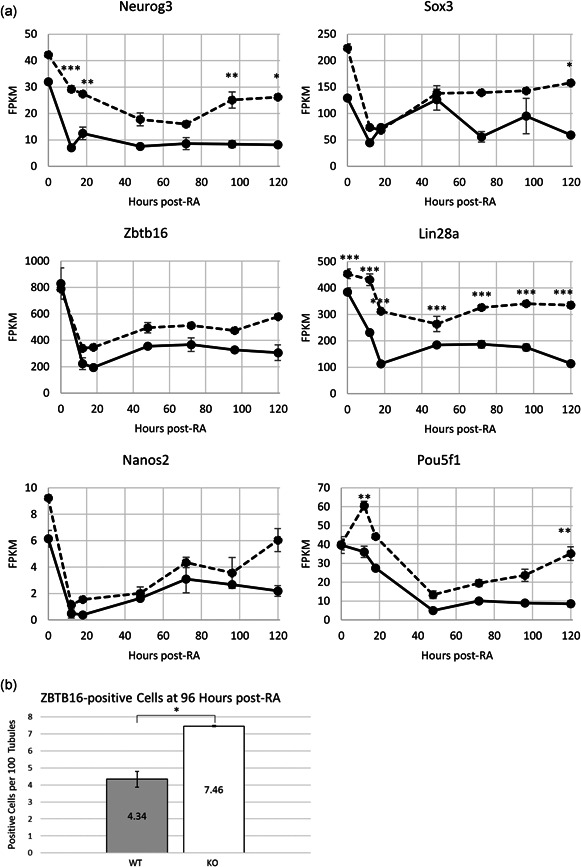
Spermatogonial development is a key process during spermatogenesis to prepare germ cells to enter meiosis. While the initial point of spermatogonial differentiation is well-characterized, the development of spermatogonia from the onset of differentiation to the point of meiotic entry has not been well defined. Further, STRA8 is highly induced at the onset of spermatogonial development but its function in spermatogonia has not been defined. To better understand how STRA8 impacts spermatogonia, we performed RNA-sequencing in both wild-type and STRA8 knockout mice at multiple timepoints during retinoic acid (RA)-stimulated spermatogonial development. As expected, in spermatogonia from wild-type mice we found that steady-state levels of many transcripts that define undifferentiated progenitor cells were decreased while transcripts that define the differentiating spermatogonia were increased as a result of the actions of RA. However, the spermatogonia from STRA8 knockout mice displayed a muted RA response such that there were more transcripts typical of undifferentiated cells and fewer transcripts typical of differentiating cells following RA action. While spermatogonia from STRA8 knockout mice can ultimately form spermatocytes that fail to complete meiosis, it appears that the defect likely begins as a result of altered messenger RNA levels during spermatogonial differentiation.

Keywords: STRA8; retinoic acid; spermatogenesis; spermatogonia; testis.


Conflict of interest statement

The authors declare that there are no conflict of interests.

Figures

Figure 1
Figure 1
(a) Experiment schematic. PIWIL2‐eGFP × STRA8 wild‐type (WT) or knockout (KO) mice were treated with WIN 18,446 daily for 7 days starting at 2 days post‐partum (dpp). Mice received a retinoic acid (RA) injection on Day 8 of treatment (9 dpp). Cells were collected via fluorescence‐activated cell sorting (FACS) at the timepoints listed in (b) to collect given spermatogonial subtypes. (b) Cell types present at given times after WIN 18,446/RA synchrony. Original data from Agrimson et al. (2016). (c) Principal component analysis (PCA) of 0‐, 12‐, 18‐, 48‐, 72‐, 96‐, and 120‐h post‐RA WT and STRA8 KO samples generated using the top 200 most variable genes. Two biological replicates at each timepoint were sequenced and nearby clustering shows high replicate similarity. k0, KO 0‐h post‐RA; k12, KO 12‐h post‐RA; k18, KO 18‐h post‐RA; k48, KO 48‐h post‐RA; k72, KO 72‐h post‐RA; k96, KO 96‐h post‐RA; k120, KO 120‐h post‐RA; w0, WT 0‐h post‐RA (Aundiff); w12, WT 12‐h post‐RA (RA‐induced genes activated); w18, WT 18‐h post‐RA (A1/A2 spermatogonia); w48, WT 48‐h post‐RA (A3 spermatogonia); w72, WT 72‐h post‐RA (A4 spermatogonia); w96, WT 96‐h post‐RA (Int spermatogonia); w120, WT 120‐h post‐RA (B spermatogonia)
Figure 2
Figure 2
(a) Numbers of genes at each timepoint for wild‐type (WT) and STRA8 knockout (KO) cells during spermatogonial development whose steady‐state levels increased or decreased relative to the corresponding Aundiff population. (b) Increased and (c) decreased expression specifically in the WT or STRA8 KO or in both the WT and KO (shared) during spermatogonial development, relative to 0‐h post‐RA (Aundiff). Blue bars show genes with increased or decreased expression specifically in the WT cells, yellow bars show genes with increased or decreased expression specifically in the STRA8 KO cells, and green bars show genes with increased or decreased expression in both the WT and STRA8 KO cells. Differentially expressed genes determined by log2 fold‐change ≥1 or ≤−1, false discovery rate ≤ 0.05
Figure 3
Figure 3
Differentially expressed genes between corresponding wild‐type (WT) and (KO) cells at 0‐ to 120‐h post‐retinoic acid (RA). Differentially expressed genes determined by R DESeq2 package (Love et al., 2014) capturing genes that had a log2 fold‐change ≥1 and false discovery rate ≤ 0.05. Gray bars represent genes that were more highly expressed in the WT cells, white bars represent genes that were more highly expressed in KO cells
Figure 4
Figure 4
(a) Pluripotency transcripts characteristic of undifferentiated spermatogonia in wild‐type (WT) and STRA8 knockout (KO) cells. Solid lines represent WT data, dashed lines represent STRA8 KO data. Data represent the average of two biological replicates, error bars represent ± SEM. Asterisks represent significance in a one‐way analysis of variance followed by a Tukey's multiple comparison honestly significant difference test, *p ≤ .05, **p ≤ .01, ***p ≤ .001. (b) ZBTB16‐positive cells at 96 h post‐retinoic acid (RA). Values represent the number of positive cells per 100 tubules, data averaged from three replicates. Asterisks show a significant difference in a paired Student's t test, two‐tailed p < .05. Gray bars show WT values, white bars show STRA8 KO values. Error bars represent ± SEM
Figure 5
Figure 5
(a) Transcripts characteristic of differentiating spermatogonia in wild‐type (WT) and STRA8 knockout (KO) cells (Kit, Esx1, and Dmrtb1). (b) Genes that have an increase in expression later during spermatogonial development (48–120 h post‐retinoic acid [RA]; Tex11 and Spag9). While there is an increase in WT expression, this increase is absent or muted in the STRA8 KO cells. (c) Mki67 transcript expression throughout spermatogonial development. Solid lines represent WT data, dashed lines represent STRA8 KO data. Data represent the average of two biological replicates, error bars represent ± SEM. Asterisks represent significance in a one‐way analysis of variance followed by a Tukey's multiple comparison honestly significant difference test, *p ≤ .05, **p ≤ .01, ***p ≤ .001
Figure 6
Figure 6
Numbers of (a) increased and (b) decreased meiotic transcripts in wild‐type (WT) and STRA8 knockout (KO) cells during spermatogonial development. Fewer differentially expressed meiotic genes were observed in STRA8 KO cells at all spermatogonial subpopulations. Differentially expressed genes determined by log2 fold‐change ≥ 1 and false discovery rate ≤ 0.05. Gray bars show differentially expressed genes compared with Aundiff in WT cells, white bars show differentially expressed genes compared with Aundiff in KO cells

References

    1. Agrimson, K. S. , Onken, J. , Mitchell, D. , Topping, T. B. , Chiarini‐Garcia, H. , Hogarth, C. A. , & Griswold, M. D. (2016). Characterizing the spermatogonial response to retinoic acid during the onset of spermatogenesis and following synchronization in the neonatal mouse testis. Biology of Reproduction, 95(4), 81. 10.1095/biolreprod.116.141770 - DOI - PMC - PubMed
    1. Anderson, E. L. , Baltus, A. E. , Roepers‐Gajadien, H. L. , Hassold, T. J. , de Rooij, D. G. , van Pelt, A. M. M. , & Page, D. C. (2008). Stra8 and its inducer, retinoic acid, regulate meiotic initiation in both spermatogenesis and oogenesis in mice. Proceedings of the National Academy of Sciences of the United States of America, 105(39), 14976–14980. 10.1073/pnas.0807297105 - DOI - PMC - PubMed
    1. Aravin, A. A. , Sachidanandam, R. , Bourc'his, D. , Schaefer, C. , Pezic, D. , Fejes Toth, K. , Bestor, T. , & Hannon, G. J. (2008). A piRNA pathway primed by individual transposons is linked to de novo DNA methylation in mice. Molecular Cell, 31(6), 785–799. 10.1016/j.molcel.2008.09.003 - DOI - PMC - PubMed
    1. Arnold, S. L. M. , Kent, T. , Hogarth, C. A. , Griswold, M. D. , Amory, J. K. , & Isoherranen, N. (2015). Pharmacological inhibition of ALDH1A in mice decreases all‐trans retinoic acid concentrations in a tissue specific manner. Biochemical Pharmacology, 95(3), 177–192. 10.1016/j.bcp.2015.03.001 - DOI - PMC - PubMed
    1. Baltus, A. E. , Menke, D. B. , Hu, Y.‐C. , Goodheart, M. L. , Carpenter, A. E. , de Rooij, D. G. , & Page, D. C. (2006). In germ cells of mouse embryonic ovaries, the decision to enter meiosis precedes premeiotic DNA replication. Nature Genetics, 38(12), 1430–1434. 10.1038/ng1919 - DOI - PubMed

Publication types