. 2021 Mar 1;94(1119):20201252.
doi: 10.1259/bjr.20201252. Epub 2021 Jan 5.

Computed tomography features of cor triatriatum: an institutional review

Affiliations

Computed tomography features of cor triatriatum: an institutional review

Archita Goel et al. Br J Radiol. .

Abstract

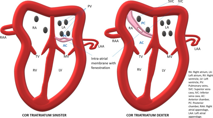
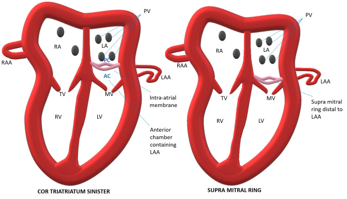
Objectives: Cor Triatriatum is a rare anomaly that can either involves the left atrium (Cor Triatriatum Sinister-CTS) or the right atrium (Cor Triatriatum Dexter- CTD). Preoperative identification of this anomaly is important in determining patient treatment course. The objective of this paper is to understand imaging findings, classification and to familiarise the reader with other associated congenital cardiac anomalies that influence patient management.

Methods: From the hospital electronic health records (EHR) database, we identified 10 patients of Cor Triatriatum out of 974 patients who underwent cardiac CT between 15 July 2014 and 20 March 2020 for congenital heart disease. Medical records and imaging findings were reviewed retrospectively.

Results: Out of 10 patients, nine patients had CTS (90%) and only one patient had CTD (10%). Five out of nine patients (55.5%) had CTS type II and four (44.4%) had CTS type III. The mean of the membrane orifices in CTS type III was 18.5 mm and was 5.78 mm in CTS type II. Pulmonary veins were dilated in all patients of CTS type II (62.5%), two patient of CTS type III (25%) and in only patient with CTD (12.5%). Ostium secundum atrial septal defect was the most common (66%) associated cardiac anomaly, followed by ventricular septal defect (44%).

Conclusions: CT allows excellent pre-operative evaluation of Cor Triatriatum and associated cardiac anomalies.

Advances in knowledge: CT is excellent in making a diagnosis and classifying Cor Triatriatum and for identification of cardiac anomalies and complications associated with it.


Conflict of interest statement

Competing interests: Nil

Figures

Figure 1.
Figure 1.
Illustration showing basic anatomy of Cor Triatriatum.
Figure 2.
Figure 2.
35-year-old female, 7-month history of dyspnoea on exertion. CT MIP images show sinus-venosus and OS-ASD (white arrow-a, c), right upper PV draining in right atrium (black arrows in a, b), fibrous membrane in right atrium from the IVC and crista terminalis to interatrial septum (curved black arrow-b, c) and dilated MPA (star-b).
Figure 3.
Figure 3.
6-year-old female with cyanosis, recurrent respiratory tract infection and failure to gain weight since birth. A common arterial trunk forming pulmonary arteries (black arrow-a) and aorta (white arrowhead-a), fibrous CTS membrane (white arrow-b) with multiple orifices (black arrowheads-b), subtruncal VSD noted (black curved arrow-c).
Figure 4.
Figure 4.
7-year-old female with 5-year history of recurrent respiratory tract infections and dyspnoea on exertion. Dilated right PVs (white arrow-a), hypoplastic left inferior PV (black arrow-a), CTS membrane (curved white arrow-b, c) proximal to left-atrial-appendage (curved black arrow-b), OS-ASD (black arrowhead-c), muscular VSDs (black arrowheads-d). Small membrane arising near mitral-annulus (white arrowhead-c). Respiratory motion-artifacts noted.
Figure 5.
Figure 5.
6-year-old female child with history of shortness of breath of 6-month duration. CT images show a large sinus venosus ASD (black arrow-a), fibrous membrane dividing left atrium (curved black arrow-a, b). Mitral valve seen separately (white arrow-a, b).
Figure 6.
Figure 6.
Illustration differentiating Cor Triatriatum Sinister from the supra mitral ring. AC, Anterior chamber; LA, Left atrium; LAA, Left atrial appendage; LV, Left ventricle; PC, Posterior chamber; PV, Pulmonary veins; RA, Right atrium; RAA, Right atrial appendage; RV, Right ventricle.

References

    1. Nassar PN, Hamdan RH. Cor triatriatum Sinistrum: classification and imaging modalities. Eur J Cardiovasc Med 2011; 1: 84–7. doi: 10.5083/ejcm.20424884.21 - DOI - PMC - PubMed
    1. Gharagozloo F, Bulkley BH, Hutchins GM. A proposed pathogenesis of cor triatriatum: impingement of the left superior vena cava on the developing left atrium. Am Heart J 1977; 94: 618–26. doi: 10.1016/S0002-8703(77)80132-4 - DOI - PubMed
    1. Simsek Z, Koza Y, Tas H. Cor triatriatum Dexter, atrial septal defects, and pulmonary stenosis-a rare association. Echocardiography 2014; 31: E124–7. doi: 10.1111/echo.12516 - DOI - PubMed
    1. Alphonso N, Nørgaard MA, Newcomb A, d'Udekem Y, Brizard CP, Cochrane A. Cor triatriatum: presentation, diagnosis and long-term surgical results. Ann Thorac Surg 2005; 80: 1666–71. doi: 10.1016/j.athoracsur.2005.04.055 - DOI - PubMed
    1. Peña E, Dennie C, Veinot J, Muñiz SH. Pulmonary hypertension: how the radiologist can help. Radiographics 2012; 32: 9–32. doi: 10.1148/rg.321105232 - DOI - PubMed

MeSH terms