

Influence of CBCT-based volumetric distortion and beam hardening artefacts on the assessment of root canal filling quality in isthmus-containing molars
- PMID: 33400563
- PMCID: PMC8231682
- DOI: 10.1259/dmfr.20200503
Influence of CBCT-based volumetric distortion and beam hardening artefacts on the assessment of root canal filling quality in isthmus-containing molars
Abstract
Objectives: To evaluate the influence of artefacts in cone beam CT (CBCT) images of filled root canals in isthmus-containing molars.
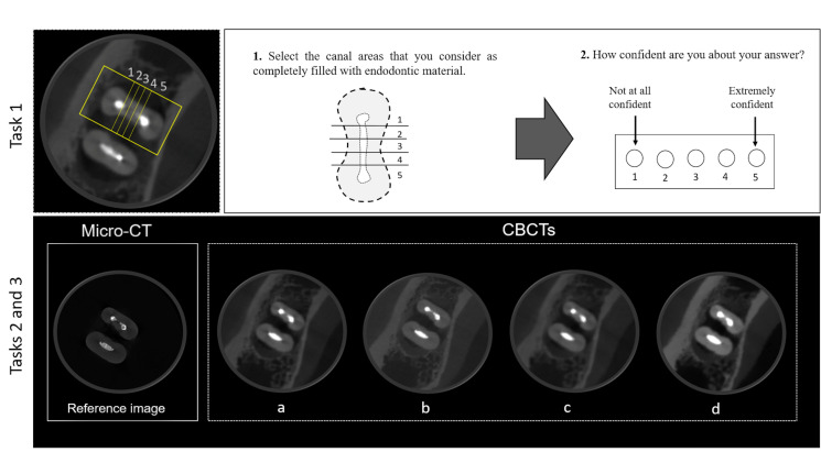
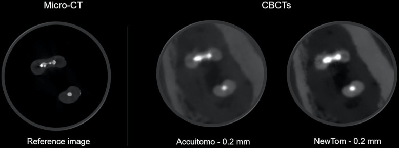
Methods: 10 teeth presenting canals with an isthmus were instrumented and filled with a thermoplasticised obturation technique. The teeth were scanned using a micro-CT device and two CBCT devices: 3D Accuitomo 170 (ACC) and NewTom VGi evo (NT), with different acquisition protocols: larger and smaller voxel size. Three examiners assessed the CBCT images for: (1) detection of filling voids; (2) assessment of under- or overestimation of the filling material and (3) resemblance of CBCT images to the reference standard. Analyses of Task 1 yielded accuracy, sensitivity and specificity for detection of filling voids. For tasks 2 and 3, statistical analysis was performed using Wilcoxon test. The level of significance was set at p < .05.
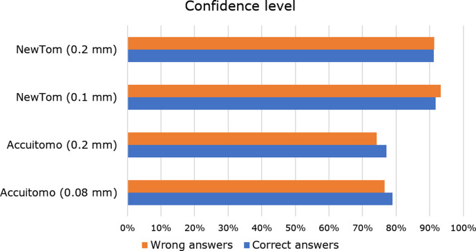
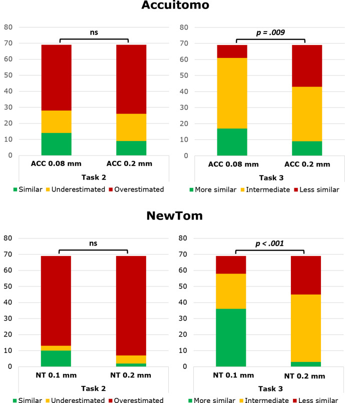
Results: For Task 1, ACC showed higher sensitivity, whereas NT presented higher specificity. No significant difference was found between the protocols in ACC, however, for NT, differences between protocols were significant for all diagnostic values. In Task 2, visualisation of the filling was overestimated for NT, while for ACC, underestimation was observed. For Task 3, images with smaller voxel size were more similar to the reference image, for both CBCT devices.
Conclusions: Different artefacts compromise the detection of filling voids on CBCT images of canals in mandibular molars with isthmus. ACC and NT present rather similar diagnostic accuracy, even though artefact expression remains device-specific.
Keywords: Artefacts; Cone-beam computed tomography; Microcomputed tomography; Root canal obturation..
Figures
References
-
- Siqueira JF, Alves FRF, Versiani MA, Rôças IN, Almeida BM, Neves MAS, et al. . Correlative bacteriologic and micro-computed tomographic analysis of mandibular molar mesial canals prepared by self-adjusting file, reciproc, and twisted file systems. J Endod 2013; 39: 1044–50. doi: 10.1016/j.joen.2013.04.034 - DOI - PubMed
MeSH terms
LinkOut - more resources
Full Text Sources
Other Literature Sources
Research Materials
