. 2020 Dec 1;11(48):4504-4520.
doi: 10.18632/oncotarget.27830.

Effect of isoform-specific HIF-1α and HIF-2α antisense oligonucleotides on tumorigenesis, inflammation and fibrosis in a hepatocellular carcinoma mouse model

Affiliations

Effect of isoform-specific HIF-1α and HIF-2α antisense oligonucleotides on tumorigenesis, inflammation and fibrosis in a hepatocellular carcinoma mouse model

Bart Vanderborght et al. Oncotarget. .

Abstract

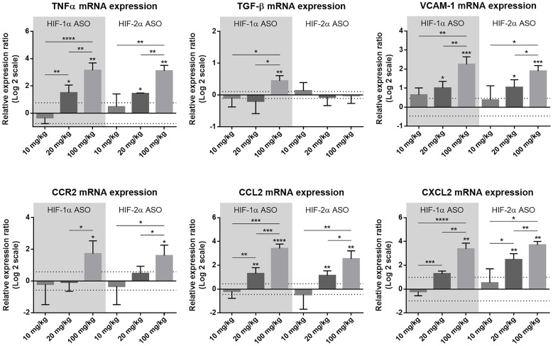
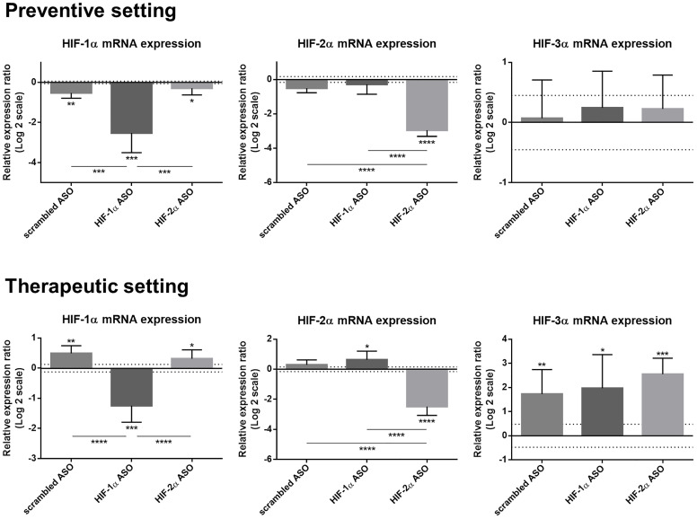
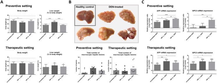
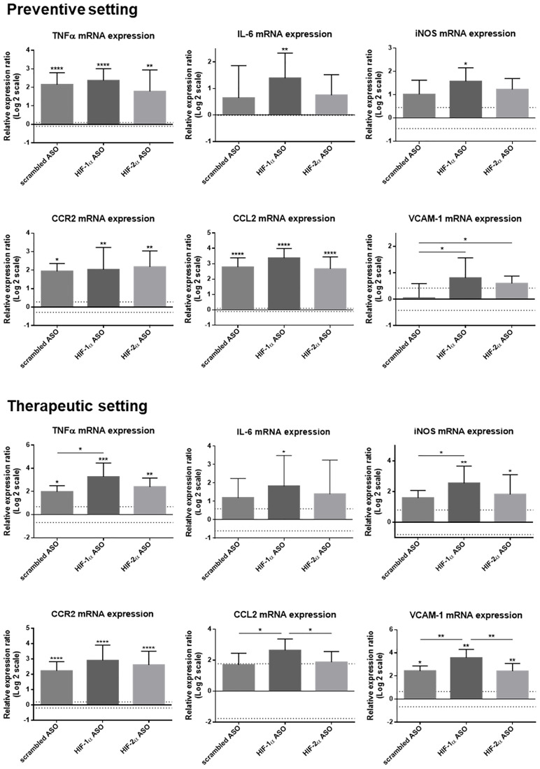
Hepatocellular carcinoma (HCC) is one of the leading causes of cancer-related death worldwide. For advanced HCC, there is still an unmet need for more effective therapeutic strategies. HCC is typically associated with hypoxia and the hypoxia-inducible factor (HIF) regulatory pathway plays an important role in HCC development and progression. Therefore, we investigated the therapeutic potential of isoform-specific HIF-1α and HIF-2α antisense oligonucleotides (ASOs), along with their effect on the inflammatory and fibrotic component of the tumor microenvironment (TME), in an experimental HCC mouse model. Based on its efficacy and safety, a dosage regimen of 20 mg/kg intraperitoneal injection of HIFα ASO twice per week was selected for further investigation in a preventive and therapeutic setting in a N,N-diethylnitrous amide (DEN)-induced HCC mouse model. DEN administration resulted in 100% tumor formation and HIFα ASO administration led to effective and selective hepatic downregulation of its target genes. HIFα ASO treatment had no effect on tumor numbers, but even enhanced the increased hepatic expression of HCC tumor markers, α-fetoprotein and glypican-3, compared to scrambled control ASO treatment in HCC mice. Especially HIF-1α ASO treatment resulted in an enhanced increase of monocytes and monocyte-derived macrophages in the liver and an enhanced hepatic upregulation of inflammatory markers. Both HIFα ASOs aggravated liver fibrosis in HCC mice compared to scrambled ASO treatment. The observed effects of our dosing regimen for HIF-1α and HIF-2α ASO treatment in the DEN-induced HCC mouse model discourage the use of HIFα isoforms as targets for the treatment of HCC.

Keywords: antisense oligonucleotides; experimental mouse model; hepatocellular carcinoma; hypoxia-inducible factor; tumor microenvironment.


Conflict of interest statement

CONFLICTS OF INTEREST The authors have no conflicts of interest to declare.

Figures

Figure 1
Figure 1. Efficacy and selectivity of different dosage regimens of HIF-1α and HIF-2α ASO.
Mice were intraperitoneally injected with 10, 20 or 100 mg/kg HIF-1α or HIF-2α ASO, or 20 mg/kg scrambled ASO, twice per week for 2 weeks. (A) Body weight and relative liver weight (expressed as % to body weight) following treatment period. The upper and lower dashed lines represent mean ± SD of the scrambled ASO data. Mean ± SD of other data are represented as bars (n = 4 per treatment group). (B) Hepatic mRNA expression of the HIFα isoforms following treatment period. The upper and lower dashed lines represent mean ± SD of the log2-transformed 20 mg/kg scrambled ASO data. Log2-transformed mean ± SD of other data, relative to the log2-transformed mean of the 20 mg/kg scrambled ASO treatment group, are represented as bars (n = 4 per treatment group). * p < 0.05, ** p < 0.01, *** p < 0.001 and **** p < 0.0001.
Figure 2
Figure 2. Effect of different dosage regimens of HIF-1α and HIF-2α ASO on hepatic expression of inflammatory markers.
Mice were intraperitoneally injected with 10, 20 or 100 mg/kg HIF-1α or HIF-2α ASO, or 20 mg/kg scrambled ASO, twice per week for 2 weeks. Hepatic mRNA expression of the inflammatory markers TNFα, TGF-β, VCAM-1, CCR2, CCL2 and CXCL2 following treatment period is shown. The upper and lower dashed lines represent mean ± SD of the log2-transformed 20 mg/kg scrambled ASO data. Log2-transformed mean ± SD of other data, relative to the log2-transformed mean of the 20 mg/kg scrambled ASO treatment group, are represented as bars (n = 4 per treatment group). * p < 0.05, ** p < 0.01, *** p < 0.001 and **** p < 0.0001.
Figure 3
Figure 3. Efficacy and selectivity of preventive and therapeutic HIF-1α and HIF-2α ASO treatment in DEN-induced HCC mice.
HCC was induced by weekly intraperitoneal DEN injection for 25 weeks. Control mice received weekly intraperitoneal 0.9% NaCl injection. DEN-treated mice were intraperitoneally injected with 20 mg/kg HIF-1α ASO, HIF-2α ASO or scrambled ASO twice per week, in either a preventive or a therapeutic setting. Control mice received scrambled ASO for the same duration of the experiment. Hepatic mRNA expression of the HIFα isoforms following preventive and therapeutic treatment is shown. The upper and lower dashed lines represent log2-transformed mean ± SD of the control mice. Bars represent log2-transformed mean ± SD of different treatment groups of DEN-treated mice, relative to the log2-transformed mean of the control mice (n = 7–9 per treatment group). * p < 0.05, ** p < 0.01, *** p < 0.001 and **** p < 0.0001.
Figure 4
Figure 4. Effect of preventive and therapeutic HIF-1α and HIF-2α ASO treatment on hepatocarcinogenesis in DEN-induced HCC mice.
HCC was induced by weekly intraperitoneal DEN injection for 25 weeks. Control mice received weekly intraperitoneal 0.9% NaCl injection. DEN-treated mice were intraperitoneally injected with 20 mg/kg HIF-1α ASO, HIF-2α ASO or scrambled ASO twice per week, in either a preventive or a therapeutic setting. Control mice received scrambled ASO for the same duration of the experiment. (A) Body weight and relative liver weight (expressed as % to body weight) following preventive and therapeutic treatment. The upper and lower dashed lines represent mean ± SD of the control mice. Bars represent mean ± SD of different treatment groups of DEN-treated mice (n = 6–8 per treatment group). (B) Upper part: Hepatic tissue of healthy control mice and DEN-treated mice. Lower part: Total number of macroscopic hepatic lesions following preventive and therapeutic treatment. Data are represented as individual values with the median (n = 6–8 per treatment group). (C) Hepatic mRNA expression of the HCC tumor markers AFP and GPC3 following preventive and therapeutic treatment. The upper and lower dashed lines represent log2-transformed mean ± SD of the control mice. Bars represent log2-transformed mean ± SD of different treatment groups of DEN-treated mice, relative to the log2-transformed mean of the control mice (n = 7–9 per treatment group). * p < 0.05, ** p < 0.01, *** p < 0.001 and **** p < 0.0001.
Figure 5
Figure 5. Effect of therapeutic HIF-1α and HIF-2α ASO treatment on hepatic macrophage pool in DEN-induced HCC mice.
HCC was induced by weekly intraperitoneal DEN injection for 25 weeks. Control mice received weekly intraperitoneal 0.9% NaCl injection. DEN-treated mice were intraperitoneally injected with 20 mg/kg HIF-1α ASO, HIF-2α ASO or scrambled ASO twice per week, in a therapeutic setting. Control mice received scrambled ASO for the same duration of the experiment. Upper part: Percentage of CD11b+Ly6C-F4/80+Tim4+ Kupffer cells, CD11b+Ly6C+F4/80+Tim4- monocytes and CD11b+Ly6C-F4/80+Tim4- monocyte-derived macrophages in live CD45+ single cell gate following treatment, measured by flow cytometry. The upper and lower dashed lines represent mean ± SD of the control mice. Bars represent mean ± SD of different treatment groups of DEN-treated mice (n = 6–7 per treatment group). * p < 0.05, ** p < 0.01, *** p < 0.001 and **** p < 0.0001. Lower part: Number of live CD45+CD11b+Ly6C-F4/80+Tim4+ Kupffer cells, CD45+CD11b+Ly6C+F4/80+Tim4- monocytes and CD45+CD11b+Ly6C-F4/80+Tim4- monocyte-derived macrophages per gram liver tissue following treatment, measured by flow cytometry. The upper and lower dashed lines represent mean ± SD of the control mice. Bars represent mean ± SD of different treatment groups of DEN-treated mice (n = 6–7 per treatment group). * p < 0.05, ** p < 0.01, *** p < 0.001 and **** p < 0.0001.
Figure 6
Figure 6. Effect of preventive and therapeutic HIF-1α and HIF-2α ASO treatment on hepatic mRNA expression of inflammatory markers in DEN-induced HCC mice.
HCC was induced by weekly intraperitoneal DEN injection for 25 weeks. Control mice received weekly intraperitoneal 0.9% NaCl injection. DEN-treated mice were intraperitoneally injected with 20 mg/kg HIF-1α ASO, HIF-2α ASO or scrambled ASO twice per week, in either a preventive or a therapeutic setting. Control mice received scrambled ASO for the same duration of the experiment. Hepatic mRNA expression of several inflammatory markers following preventive and therapeutic treatment is shown. The upper and lower dashed lines represent log2-transformed mean ± SD of the control mice. Bars represent log2-transformed mean ± SD of different treatment groups of DEN-treated mice, relative to the log2-transformed mean of the control mice (n = 7–9 per treatment group). * p < 0.05, ** p < 0.01, *** p < 0.001 and **** p < 0.0001.
Figure 7
Figure 7. Effect of preventive and therapeutic HIF-1α and HIF-2α ASO treatment on hepatic protein expression of inflammatory markers in DEN-induced HCC mice.
HCC was induced by weekly intraperitoneal DEN injection for 25 weeks. Control mice received weekly intraperitoneal 0.9% NaCl injection. DEN-treated mice were intraperitoneally injected with 20 mg/kg HIF-1α ASO, HIF-2α ASO or scrambled ASO twice per week, in either a preventive or a therapeutic setting. Control mice received scrambled ASO for the same duration of the experiment. Hepatic protein expression of several inflammatory markers following preventive and therapeutic treatment is shown. The upper and lower dashed lines represent mean ± SD of the control mice. Bars represent mean ± SD of different treatment groups of DEN-treated mice (n = 7–9 per treatment group). * p < 0.05, ** p < 0.01, *** p < 0.001 and **** p < 0.0001.
Figure 8
Figure 8. Effect of preventive and therapeutic HIF-1α and HIF-2α ASO treatment on liver fibrosis in DEN-induced HCC mice.
HCC was induced by weekly intraperitoneal DEN injection for 25 weeks. Control mice received weekly intraperitoneal 0.9% NaCl injection. DEN-treated mice were intraperitoneally injected with 20 mg/kg HIF-1α ASO, HIF-2α ASO or scrambled ASO twice per week, in either a preventive or a therapeutic setting. Control mice received scrambled ASO for the same duration of the experiment. Upper part: Representative histological images of Sirius Red-stained liver sections with Metavir fibrosis stage F0 (no fibrosis), F1 (portal fibrosis without septa) and F2 (portal fibrosis with few septa). Magnification 200×. Lower part: Degree of hepatic fibrosis following preventive and therapeutic treatment, assessed through histological analysis of Sirius Red-stained liver sections using the Metavir scoring system. Data are represented as individual values with the median (n = 7–9 per HCC treatment group).
Figure 9
Figure 9. Effect of preventive and therapeutic HIF-1α and HIF-2α ASO treatment on fibrotic markers in the liver of DEN-induced HCC mice.
HCC was induced by weekly intraperitoneal DEN injection for 25 weeks. Control mice received weekly intraperitoneal 0.9% NaCl injection. DEN-treated mice were intraperitoneally injected with 20 mg/kg HIF-1α ASO, HIF-2α ASO or scrambled ASO twice per week, in either a preventive or a therapeutic setting. Control mice received scrambled ASO for the same duration of the experiment. Hepatic mRNA expression of fibrotic markers following preventive and therapeutic treatment is shown. The upper and lower dashed lines represent log2-transformed mean ± SD of the control mice. Bars represent log2-transformed mean ± SD of different treatment groups of DEN-treated mice, relative to the log2-transformed mean of the control mice (n = 7–9 per treatment group). * p < 0.05, ** p < 0.01, *** p < 0.001 and **** p < 0.0001.
Figure 10
Figure 10. Preventive and therapeutic treatment of ASOs in experimental HCC.
HCC was induced by weekly intraperitoneal DEN injection for 25 weeks. Control mice received weekly intraperitoneal 0.9% NaCl injection. DEN-treated mice were intraperitoneally injected with 20 mg/kg HIF-1α ASO, HIF-2α ASO or scrambled ASO twice per week. In the preventive setting, ASO injections started 10 weeks following the first DEN injection (at 15 weeks of age) and persisted for a period of 15 weeks. In the therapeutic setting, ASO injections started 20 weeks following the first DEN injection (at 25 weeks of age) and persisted for a period of 8 weeks. Control mice received scrambled ASO for the same duration of the experiment. Following respective treatment periods, mice were sacrificed and the liver was isolated for further analyses.

References

    1. Yang JD, Hainaut P, Gores GJ, Amadou A, Plymoth A, Roberts LR. A global view of hepatocellular carcinoma: trends, risk, prevention and management. Nat Rev Gastroenterol Hepatol. 2019; 16:589–604. 10.1038/s41575-019-0186-y. - DOI - PMC - PubMed
    1. Villanueva A. Hepatocellular Carcinoma. N Engl J Med. 2019; 380:1450–62. 10.1056/NEJMra1713263. - DOI - PubMed
    1. Balogh J, Victor D 3rd, Asham EH, Burroughs SG, Boktour M, Saharia A, Li X, Ghobrial RM, Monsour HP Jr. Hepatocellular carcinoma: a review. J Hepatocell Carcinoma. 2016; 3:41–53. 10.2147/JHC.S61146. - DOI - PMC - PubMed
    1. Dhanasekaran R, Bandoh S, Roberts LR. Molecular pathogenesis of hepatocellular carcinoma and impact of therapeutic advances. F1000Res. 2016; 5:F1000 Faculty Rev–879. 10.12688/f1000research.6946.1. - DOI - PMC - PubMed
    1. Forner A, Reig M, Varela M, Burrel M, Feliu J, Briceño J, Sastre J, Martí-Bonmati L, Llovet JM, Bilbao JI, Sangro B, Pardo F, Ayuso C, et al. [Diagnosis and treatment of hepatocellular carcinoma. Update consensus document from the AEEH, SEOM, SERAM, SERVEI and SETH]. Med Clin (Barc). 2016; 146:511.e1–511.e22. 10.1016/j.medcli.2016.01.028. - DOI - PubMed

LinkOut - more resources