Review
. 2021 Mar:170:83-112.
doi: 10.1016/j.addr.2020.12.014. Epub 2021 Jan 2.

Self-assembled mRNA vaccines

Affiliations
Review

Self-assembled mRNA vaccines

Jeonghwan Kim et al. Adv Drug Deliv Rev. 2021 Mar.

Abstract

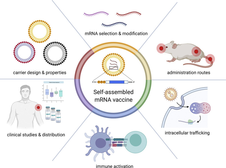
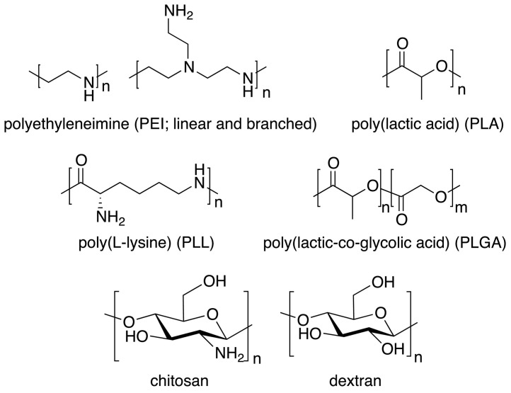
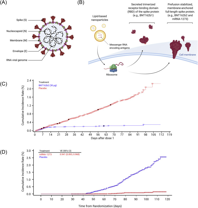
mRNA vaccines have evolved from being a mere curiosity to emerging as COVID-19 vaccine front-runners. Recent advancements in the field of RNA technology, vaccinology, and nanotechnology have generated interest in delivering safe and effective mRNA therapeutics. In this review, we discuss design and self-assembly of mRNA vaccines. Self-assembly, a spontaneous organization of individual molecules, allows for design of nanoparticles with customizable properties. We highlight the materials commonly utilized to deliver mRNA, their physicochemical characteristics, and other relevant considerations, such as mRNA optimization, routes of administration, cellular fate, and immune activation, that are important for successful mRNA vaccination. We also examine the COVID-19 mRNA vaccines currently in clinical trials. mRNA vaccines are ready for the clinic, showing tremendous promise in the COVID-19 vaccine race, and have pushed the boundaries of gene therapy.

Keywords: COVID-19; Gene delivery; Immunization; Lipid nanoparticles; Self-assembly; mRNA delivery.


Conflict of interest statement

Conflicts of Interest There are no conflicts to declare.

Figures

Unlabelled Image
Graphical abstract
Fig. 1
Fig. 1
(A, B) Schematic diagrams of in-vitro-transcribed mRNA: (A) non-replicating mRNA and (B) self-amplifying RNA. (C) Replication of self-amplifying RNA inside cells. (1) After translation, non-structural proteins 1-4 (nsP1-4) are produced, forming the early replication complex that synthesizes (-) strand of mRNA transcript. (2, 3) The late replication complex produces (2) genomic (+) strand and (3) subgenomic (+) strand. Genomic transcript continues further replications and subgenomic transcript initiates antigen production.
Fig. 2
Fig. 2
Modes of action of intramuscularly administered mRNA vaccines. (1, 2) mRNA vaccines can transfect (1) muscle cells as well as (2) tissue-resident APCs near the injection site. (3) mRNA vaccines can flow into proximal lymph nodes (LNs) and transfect LN-resident cells, resulting in activation of T and B cell development.
Fig. 3
Fig. 3
Mechanism of adaptive immune responses induced by mRNA vaccines. (1) Endosomal escape of mRNA to the cytosol after endocytosis-mediated internalization. (2) Antigen protein translated from exogenous mRNA is degraded into fragments in proteasome, followed by MHC-I presentation. (3) Antigen protein can undergo lysosomal degradation via various mechanisms, such as autophagy and signal peptide, followed by MHC-II presentation. (4) Antigen protein can be destined to extracellularly express in secreted or membrane-anchored form. (5) Extracellularly expressed antigen can be taken up again by APCs, directed to lysosomal degradation. (6) Instead, the extracellular antigen can be recognized by B cell receptor on B cells, leading to B cell maturation. (7) MHC-I presents the epitope to CD8+ T cells whereas (8) MHC-II presents the epitope to CD4+ T cells.
Fig. 4
Fig. 4
Hypothetical mechanisms of endosomal escape of nanocarriers. (A) Nanocarriers can induce destabilization of endosomal membrane for cytosolic release of genetic cargos. (B) Nanocarriers, particularly polyplexes, can scavenge protons and become cationic in acidic lumens of endosome compartments, resulting in the inflow of more protons and counter ions. This osmotic gradient induces influx of water to the endosomes, causing endosome rupture. (C) Nanocarriers swell in acidic pH due to the electrostatic repulsion and physically rupture the endosome. Reproduced from [121] with permission.
Fig. 5
Fig. 5
Non-covalent interactions in supramolecular chemistry. Electrostatic interactions determine the encapsulation of mRNA and endosomal escape while hydrophobic interactions likely affect the formation and long-term stability of the delivery vector. The roles of other interactions in self-assembly of mRNA vaccines are not yet understood. Adapted with permission from [135].
Fig. 6
Fig. 6
(A) General structure of lipid nanoparticles. Ionizable or cationic lipids (yellow box) are the main component responsible for the encapsulation of nucleic acid and intracellular delivery. These lipids may be divided into groups shown below. (B) Structures of first-generation cationic lipids DOTMA, DOTAP, and DOGS; (C) Structures of ionizable lipids DODMA and DLin-MC3-DMA; (D) Structures of selected ester-based and disulfide-based biodegradable ionizable lipids; (E) Structures of selected ionizable lipidoids.
Fig. 7
Fig. 7
Rational development of Dlin-MC3-DMA (MC3) from DODMA.
Fig. 8
Fig. 8
Selected structures of natural and synthetic polymers used for mRNA complexation.
Fig. 9
Fig. 9
(A) Structure of SARS-CoV-2 virus depicting a RNA genome and structural proteins [nucleocapsid (N), membrane (M), envelope (E) proteins, and spike (S) protein], (non-structural proteins are not shown). (B) Pfizer/BioNTech (BNT162b2) and Moderna (mRNA-1273) COVID-19 vaccines both utilize LNP platform and carry mRNA encoding the prefusion-stabilized, membrane-anchored, full-length spike protein of SARS-CoV-2. BNT162b1, another vaccine from Pfizer/BioNTech, is a LNP-formulated mRNA vaccine encoding the secreted trimerized RBD of the SARS-CoV-2 spike protein. (C) Cumulative incidence curves for the first COVID-19 occurrence after dose 1 of BNT162b2 vaccine. Reprinted from [301]. (D) Cumulative incidence curves for the first COVID-19 occurrence after randomization (same as date of dose 1) of mRNA-1273 vaccine. Reprinted from [305].

References

    1. Sahin U., Karikó K., Türeci Ö. mRNA-based therapeutics — developing a new class of drugs. Nat. Rev. Drug Discov. 2014;13:759–780. doi: 10.1038/nrd4278. - DOI - PubMed
    1. Pardi N., Hogan M.J., Porter F.W., Weissman D. mRNA vaccines — a new era in vaccinology. Nat. Rev. Drug Discov. 2018;17:261–279. doi: 10.1038/nrd.2017.243. - DOI - PMC - PubMed
    1. Granot Y., Peer D. Delivering the right message: Challenges and opportunities in lipid nanoparticles-mediated modified mRNA therapeutics—An innate immune system standpoint. Interact. Nanoparticles Immune Syst. Advant. Disadv. 2017;34:68–77. doi: 10.1016/j.smim.2017.08.015. - DOI - PubMed
    1. Kowalski P.S., Rudra A., Miao L., Anderson D.G. Delivering the messenger: advances in technologies for therapeutic mRNA delivery. Mol. Ther. 2019;27:710–728. doi: 10.1016/j.ymthe.2019.02.012. - DOI - PMC - PubMed
    1. Wang Y., Yu C. Emerging concepts of nanobiotechnology in mRNA delivery. Angew. Chem. Int. Ed. 2020 doi: 10.1002/anie.202003545. - DOI - PubMed

Publication types

MeSH terms