Review
. 2021 Jan 1;12(1):45.
doi: 10.3390/mi12010045.

Engineered Microgels-Their Manufacturing and Biomedical Applications

Affiliations
Review

Engineered Microgels-Their Manufacturing and Biomedical Applications

Hamzah Alzanbaki et al. Micromachines (Basel). .

Abstract

Microgels are hydrogel particles with diameters in the micrometer scale that can be fabricated in different shapes and sizes. Microgels are increasingly used for biomedical applications and for biofabrication due to their interesting features, such as injectability, modularity, porosity and tunability in respect to size, shape and mechanical properties. Fabrication methods of microgels are divided into two categories, following a top-down or bottom-up approach. Each approach has its own advantages and disadvantages and requires certain sets of materials and equipments. In this review, we discuss fabrication methods of both top-down and bottom-up approaches and point to their advantages as well as their limitations, with more focus on the bottom-up approaches. In addition, the use of microgels for a variety of biomedical applications will be discussed, including microgels for the delivery of therapeutic agents and microgels as cell carriers for the fabrication of 3D bioprinted cell-laden constructs. Microgels made from well-defined synthetic materials with a focus on rationally designed ultrashort peptides are also discussed, because they have been demonstrated to serve as an attractive alternative to much less defined naturally derived materials. Here, we will emphasize the potential and properties of ultrashort self-assembling peptides related to microgels.

Keywords: 3D bioprinting; biofabrication; cell-laden constructs; microgels; self-assembling peptides.


Conflict of interest statement

The authors declare no conflict of interest.

Figures

Figure 1
Figure 1
Microfluidic emulsion set up. The two immiscible fluids (water and oil) are pumped out of the syringes into the microfluidic chip. The relative flowrates of the two fluids, as well as the geometry of the microfluidic chip, dictate the size of the droplets generated in the chip. The droplets transform into microgels, which are then collected and washed to remove excess oil.
Figure 2
Figure 2
(A) The centrifugal microfluidic device consisting of a microtube, a Theta capillary holding the alginate solution, a capillary holder and calcium chloride solution. When centrifugal force is applied, droplets of the alginate solution fall into the CaCl2 solution, where gelation takes place. (B) The in-air microfluidic working principle is shown here as two jets of two reactive liquids that meet to form droplets which are then transformed to microgels. Upon the contact of the two jets, gelation begins, and the droplet transforms into a microgel particle. Panel (A) has been reproduced with a permission from Wiley [59] and Science Advances for Panel (B) [58].
Figure 3
Figure 3
Fabrication of heparin microgels and their loading with human bone morphogenetic protein-2. Heparin methacrylamide was used to fabricate the microgels in a water-in-oil emulsion. APS/TEMED refers to the free radical initiators ammonium persulfate (APS) and tetramethylethane-1,2-diamine (TEMED), respectively. HNSO3− and SO4− are the sulfate groups on heparin that bind to BMP-2. The figure has been reproduced with permission from Science Advances [62].
Figure 4
Figure 4
Fabrication of mesenchymal stromal cells (MSC) cell-laden chitosan-collagen microgels. Emulsification was used to generate the microgels with a particle diameter of about 100 µm. The microgels were able to maintain cell viability for up to 21 days. The figure has been reproduced with permission from Elsevier [74].
Figure 5
Figure 5
Design and working principle of a biocompatible device housing therapeutic cells. The cells are embedded in alginate microgels allowing for the intake of nutrients and oxygen, removal of waste and secretion of therapeutic proteins while keeping the host’s immune cells from entering the device. The figure has been reproduced with permission from Springer Nature [78].
Figure 6
Figure 6
Setup of 3D bioprinting of PEG microgels. The extrusion of the microgels requires low applied force, allowing for cell incorporation while maintaining cell viability. Crosslinking the microgels via thiol-ene reaction allows for long-term stability. The figure has been reproduced with permission from the Royal Society of Chemistry [84].
Figure 7
Figure 7
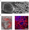
(A) SEM image of a single microgel (left) showing the round spherical shape and the network of fibers (right) resulting from the self-assembling of the ultrashort peptides. The figures have been adopted from [108]. (B) Vascularization of 3D bulk hydrogel with embedded endothelial cell-laden microgels. Anti-CD31 Alexa Fluor (red) was used for the detection of endothelial cells. 4′,6-diamidino-2-phenylindole (DAPI) (blue) was used for the detection of nuclei. The right image is a zoom-in of the left image, and both images show that the vascular network originated from the embedded microgels and spread to the rest of the bulk hydrogel.
Figure 8
Figure 8
Principle and working mechanism of the self-assembling therapeutic peptide diCPT-iRGD. The resulting hydrogel releases camptothecin (CPT) when reacting with glutathione (GSH) and releases c-di-AMP (CDA) as the hydrogel degrades. This stimulates the STING pathway and leads to the infiltration of activated immune cells, thus leading to the elimination of cancerous cells. The figure has been reproduced with permission from Springer Nature [113].

References

    1. Dai Z., Ngai T. Microgel particles: The structure-property relationships and their biomedical applications. J. Polym. Sci. Part A Polym. Chem. 2013;51:2995–3003. doi: 10.1002/pola.26698. - DOI
    1. Lee K.Y., Mooney D.J. Alginate: Properties and biomedical applications. Prog. Polym. Sci. 2012;37:106–126. doi: 10.1016/j.progpolymsci.2011.06.003. - DOI - PMC - PubMed
    1. Daly A.C., Riley L., Segura T., Burdick J.A. Hydrogel microparticles for biomedical applications. Nat. Rev. Mater. 2019;5:20–43. doi: 10.1038/s41578-019-0148-6. - DOI - PMC - PubMed
    1. Bohn P., Meier M.A.R. Uniform poly(ethylene glycol): A comparative study. Polym. J. 2020;52:165–178. doi: 10.1038/s41428-019-0277-1. - DOI
    1. Munim S.A., Raza Z.A. Poly(lactic acid) based hydrogels: Formation, characteristics and biomedical applications. J. Porous Mat. 2019;26:881–901. doi: 10.1007/s10934-018-0687-z. - DOI