. 2021 Jan 1;10(1):58.
doi: 10.3390/cells10010058.

Human Adipose Tissue-Derived Mesenchymal Stromal Cells Inhibit CD4+ T Cell Proliferation and Induce Regulatory T Cells as Well as CD127 Expression on CD4+CD25+ T Cells

Affiliations

Human Adipose Tissue-Derived Mesenchymal Stromal Cells Inhibit CD4+ T Cell Proliferation and Induce Regulatory T Cells as Well as CD127 Expression on CD4+CD25+ T Cells

Agnese Fiori et al. Cells. .

Abstract

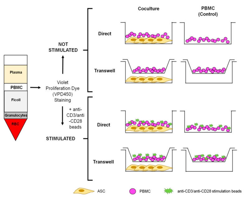
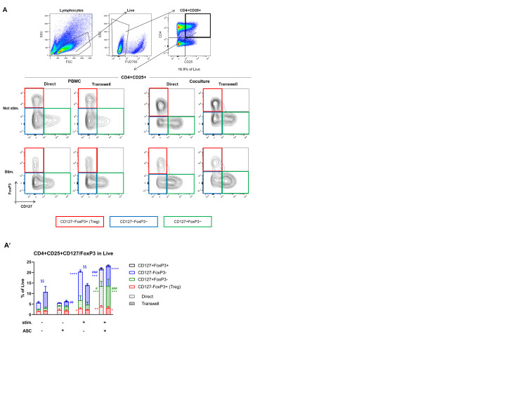
Mesenchymal stromal cells (MSC) exert their immunomodulatory potential on several cell types of the immune system, affecting and influencing the immune response. MSC efficiently inhibit T cell proliferation, reduce the secretion of pro-inflammatory cytokines, limit the differentiation of pro-inflammatory Th subtypes and promote the induction of regulatory T cells (Treg). In this study, we analyzed the immunomodulatory potential of human adipose tissue-derived MSC (ASC), on CD4+ T cells, addressing potential cell-contact dependency in relation to T cell receptor stimulation of whole human peripheral blood mononuclear cells (PBMC). ASC were cultured with not stimulated or anti-CD3/CD28-stimulated PBMC in direct and transwell cocultures; PBMC alone were used as controls. After 7 days, cocultures were harvested and we analyzed: (1) the inhibitory potential of ASC on CD4+ cell proliferation and (2) phenotypic changes in CD4+ cells in respect of Treg marker (CD25, CD127 and FoxP3) expression. We confirmed the inhibitory potential of ASC on CD4+ cell proliferation, which occurs upon PBMC stimulation and is mediated by indoleamine 2,3-dioxygenase. Importantly, ASC reduce both pro- and anti-inflammatory cytokine secretion, without indications on specific Th differentiation. We found that stimulation induces CD25 expression on CD4+ cells and that, despite inhibiting overall CD4+ cell proliferation, ASC can specifically induce the proliferation of CD4+CD25+ cells. We observed that ASC induce Treg (CD4+CD25+CD127-FoxP3+) only in not stimulated cocultures and that ASC increase the ratio of CD4+CD25+CD127+FoxP3- cells at the expense of CD4+CD25+CD127-FoxP3- cells. Our study provides new insights on the interplay between ASC and CD4+ T cells, proposing that ASC-dependent induction of Treg depends on PBMC activation which affects the balance between the different subpopulations of CD4+CD25+ cells expressing CD127 and/or FoxP3.

Keywords: CD127; CD4 T cells; immunomodulation; mesenchymal stromal cells; regulatory T cells.


Conflict of interest statement

The authors declare no conflict of interest.

Figures

Figure 1
Figure 1
Schematic representation of the experimental conditions. Stimulated and not stimulated PBMC are seeded with or without ASC in direct and transwell cultures. PBMC monocultures represent the control conditions, respectively. ASC: adipose tissue-derived stromal cells; PBMC: peripheral blood mononuclear cells; RBC: red blood cells.
Figure 2
Figure 2
Adipose tissue-derived mesenchymal stromal cells (ASC) inhibit CD4+ cell proliferation. (A) Quantitative assessment of CD4+ cell proliferation using the Proliferation tool (FlowJo 7). First, the peak zero is set based on the VPD450 intensity in non-proliferated cells (orange peak). Then, the proliferation tool algorithm calculates the series of generations (pink peaks) and the division index (DI) to quantify proliferation. Dashed line: first generation peak. (A′) The DI of CD4+ within stimulated PBMC is significantly increased compared to controls (DI set to 1; direct: p < 0.0001, transwell: p < 0.01). ASC significantly reduce the DI in cocultures with stimulated PBMC (direct: p < 0.001, transwell: p < 0.01). n = 5, 2-way ANOVA multiple comparisons. (B) At day 7, indoleamine 2,3-dioxygenase (IDO) levels are significantly higher in ASC of stimulated cocultures compared to ASC of not stimulated cocultures (direct: p < 0.0001, transwell: p < 0.01). IDO was not detectable in ASC monoculture. n = 4, 2-way ANOVA multiple comparisons. (B′) At day 7, kynurenine levels in conditioned media (CM) of cocultures are higher in direct than in transwell not stimulated cocultures (p < 0.01) and significantly increased in the stimulated setting (direct: p < 0.01, transwell p < 0.001). Kynurenine was not detected in any monoculture CM. n = 4, 2-way ANOVA multiple comparisons.
Figure 3
Figure 3
Cocultures reduce Th1/Th2 cytokine secretion and have increased levels of transforming growth factor beta (TGF-β). (A) Upon stimulation, interleukin (IL)-5 concentration is higher in direct PBMC (p < 0.01; direct vs. transwell p < 0.05) and direct cocultures (p < 0.05) compared to their controls. (B) Levels of IL-9 are more concentrated in stimulated direct PBMC than not stimulated PBMC (p < 0.01) and stimulated cocultures (p < 0.01). In not stimulated direct cocultures, IL-9 was not detectable (n.d.). (C) Upon stimulation, IL-10 is more concentrated in CM of PBMC than in CM of its controls (at least p < 0.05) and stimulated cocultures (transwell p < 0.01). (D) IL-13 concentration is higher in stimulated direct PBMC than not stimulated PBMC (p < 0.05) and direct stimulated cocultures (p < 0.05). IL-13 was not detectable in not stimulated direct cocultures. (E) IL-17a is significantly higher concentrated in CM of direct stimulated PBMC (direct vs. transwell p < 0.01) than not stimulated PBMC (p < 0.001) and stimulated cocultures (p < 0.001). (F) IL-17f is more concentrated in CM of stimulated direct PBMC (direct vs. transwell, p < 0.01) than not stimulated PBMC (p < 0.001) and stimulated cocultures (p < 0.001). (G) IL-22 concentration is higher in stimulated direct PBMC (direct vs. transwell, p < 0.05) than in CM of not stimulated PBMC (p < 0.001) and stimulated cocultures (p < 0.01). (H) TNF-α (tumor necrosis factor alpha) is significantly higher concentrated in CM of PBMC upon stimulation and presence of ASC (at least p < 0.05). TNF-α was not detectable in not stimulated direct cocultures. (I) Cocultures show significant higher levels of TGF-β compared to monocultures (at least p < 0.05). Levels are significantly higher in not stimulated condition (p < 0.001) and in direct coculture (p < 0.0001). n.d. = not detectable. n = 4, 2-way ANOVA multiple comparisons.
Figure 4
Figure 4
ASC increase CD25+ cells within the CD4+ compartment upon stimulation. (A) Representative gating strategy for CD4+CD25+ cells. Lymphocytes are gated based on sideward scatter (SSC) and forward scatter (FSC), then live cells are gated (FVD780 negative). Then, CD4+CD25+ percentage is calculated referring to the live population. One representative experiment is shown. (A′) Upon stimulation, CD25 expression is significantly increased (at least p < 0.05) and further increased by ASC cocultures (p < 0.05 in transwell). n = 3, 2-way ANOVA multiple comparison. (B) Representative gating strategy for proliferated and not proliferated CD4+CD25+ cells. Lymphocytes are gated based on SSC and FSC, and then live cells are gated (FVD780 negative). Proliferated and not proliferated cells are gated based on VPD450 intensity. Frequencies of CD4+CD25+ are then calculated within proliferated/not proliferated populations. One representative experiment shown. (B′) The combination of stimulation and cocultures induce significant CD4+CD25+ proliferation compared to the respective controls (at least p < 0.001). Contrarily, in not stimulated conditions, cocultures result in a significantly reduced proportion of proliferated CD4+CD25+ (at least p < 0.01 and direct vs. transwell p < 0.01). (B′′) Upon stimulation, the proportion of not proliferated CD4+CD25+ is significantly increased irrespective of ASC (at least p < 0.05). n = 3, 2-way ANOVA multiple comparison.
Figure 5
Figure 5
ASC induce regulatory T cells (Treg) and CD127+FoxP3- upon stimulation. (A) Representative gating strategy for Treg. Lymphocytes are gated based on SSC and FSC, then live cells are gated (FVD780 negative). Then, CD4/CD25 double-positive cells are gated and investigated for CD127 and FoxP3 expression. Red gate: CD127-FoxP3+ cells (Treg), blue gate CD127-FoxP3-, green gate CD127+FoxP3-. One representative experiment shown. (A′) Stimulation induces a slight increase in Treg (p < 0.05 in direct) which is further increased by ASC (at least p < 0.05). CD127+FoxP3- cells are increased in cocultures (not significant) but to a much higher extent in stimulated cocultures (at least p < 0.05). The percentage of CD127-FoxP3- cells is significantly reduced by ASC in both stimulated and not stimulated conditions (at least p < 0.01) with respect to their controls. Percentages are calculated referring to the starting live population. n = 3, 2-way ANOVA multiple comparison.
Figure 6
Figure 6
ASC significantly change the proportion of Treg and CD127+FoxP3- cells within the proliferated/not proliferated CD4+CD25+ subgroup. (A) The proportion of Treg within proliferated CD4+CD25+ cells is significantly increased in not stimulated cocultures (p < 0.001) but reduced in stimulated cocultures (p < 0.05). The percentage of CD127-FoxP3- cells within proliferated CD4+CD25+ cells is significantly lower in cocultures compared to their respective controls (at least p < 0.01) while the percentage of CD127+FoxP3- is higher (at least p < 0.01). (A′). Within the not proliferated CD4+CD25+ subgroup, the proportion of Treg is significantly reduced upon stimulation irrespective of cocultures (at least p < 0.05). The proportion of CD127+FoxP3- in the not proliferated cells is significantly increased in presence of ASC (at least p < 0.001). n = 3, 2-way ANOVA multiple comparison.

Similar articles

Cited by

References

    1. González M.A., Gonzalez-Rey E., Rico L., Büscher D., Delgado M. Adipose-derived mesenchymal stem cells alleviate experimental colitis by inhibiting inflammatory and autoimmune responses. Gastroenterology. 2009;136:978–989. doi: 10.1053/j.gastro.2008.11.041. - DOI - PubMed
    1. Gonzalez-Rey E., Gonzalez M.A., Varela N., O’Valle F., Hernandez-Cortes P., Rico L., Büscher D., Delgado M. Human adipose-derived mesenchymal stem cells reduce inflammatory and T cell responses and induce regulatory T cells in vitro in rheumatoid arthritis. Ann. Rheum. Dis. 2010;69:241. doi: 10.1136/ard.2008.101881. - DOI - PubMed
    1. Yan Z., Zhuansun Y., Liu G., Chen R., Li J., Ran P. Mesenchymal stem cells suppress T cells by inducing apoptosis and through PD-1/B7-H1 interactions. Immunol. Lett. 2014;162:248–255. doi: 10.1016/j.imlet.2014.09.013. - DOI - PubMed
    1. Di Nicola M., Carlo-Stella C., Magni M., Milanesi M., Longoni P.D., Matteucci P., Grisanti S., Gianni A.M. Human bone marrow stromal cells suppress T-lymphocyte proliferation induced by cellular or nonspecific mitogenic stimuli. Blood. 2002;99:6. doi: 10.1182/blood.V99.10.3838. - DOI - PubMed
    1. Tse W.T., Pendleton J.D., Beyer W.M., Egalka M.C., Guinan E.C. Suppression of allogeneic T-cell proliferation by human marrow stromal cells: Implications in transplantation. Transplantation. 2003;75:389–397. doi: 10.1097/01.TP.0000045055.63901.A9. - DOI - PubMed

Publication types

MeSH terms